The computed tomographyalso known as computed axial tomography, also known as computed tomography, or TAChas become one of the most popular techniques of image diagnosis most commonly used. It is a procedure that uses special X-ray equipment and advanced computers to obtain three-dimensional images with different slices of the body.
Since its clinical introduction in 1971, it has undergone successive advances that have allowed its application in different fields of medicine. At present, computed tomography is used to diagnose disorders such as cancer, cardiovascular conditions, infectious processes, trauma and diseases of the locomotor apparatus. In the following article, we analyze how it works, what it is used for and the origin and evolution of this diagnostic test.
How does a CAT scan work?
In order to perform this diagnostic imaging, the following is used computed axial tomography system which incorporates a X-ray scanners generating three-dimensional images with different cuts of the interior of the organism.
These slices are called tomographic images and are used for the following purposes study various internal regions of the bodyThe CT scanner can be used to view everything from organs, bones and soft tissues to blood vessels. In contrast to radiography, which only provides a two-dimensional representation, the CT scan makes it possible to observe the three-dimensional images. This makes it possible to analyze tissues with greater detail and clarity. Another aspect to note is that the CT scanner utilizes a X-ray source and has a ionizing radiation higher than that of an X-ray.
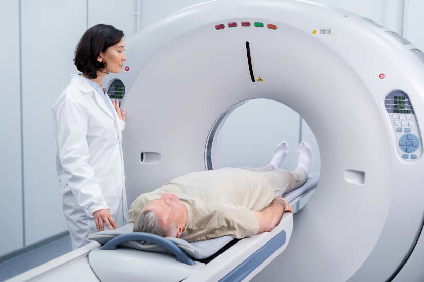
During the procedure, the CT scanner rotates around the circular opening of a threaded structure called Gantry. The patient lies on a bed and is inserted inside the scanner so that the specialist can analyze the tissues. The X-ray detectors are located in front of the X-ray source and generate a series of images through different cuts. Subsequently, are transmitted to a computer where the interior of the organism can be visualized and analyzed.
CT contrast medium
As with X-rays, dense structures within the body, such as bones, are easy to image. However, soft tissues are more difficult to image. For this reason, contrast media have been developed that increase the visibility of tissues during X-ray or CT scans. Contain a set of substances that are safe for patients and allow the X-rays to be stopped, so that the organs will be seen in greater detail in the test.
For example, to examine the circulatory system, an iodine-based intravenous contrast medium is injected into the bloodstream to illuminate the blood vessels.
What is CT used for?
CT is used as a clinical diagnostic test, in follow-up studies to analyze the patient's health status, in radiotherapy treatment planning, and even for screening asymptomatic individuals with specific risk factors. A computed tomography scan creates detailed images of the bodywhich include the brain, thorax, spine and abdomen.. Specifically, we can highlight the following uses:
- To help diagnose the presence of a cancer or tumor.. It is one of the most widely used techniques to screen for the presence of colorectal cancer and lung cancer.
- Obtain information about the stage of a cancer.
- Determine if a cancer reacts to treatment.
- To detect the return or recurrence of a tumor.
- Diagnose an infection.
- Support technique to guide a biopsy procedure.
- To guide some local treatmentssuch as cryotherapy, radiofrequency ablation and radioactive seed implantation.
- Radiotherapy planning external beam or surgery.
- Study the blood vessels.
When did computed tomography come into being?
Computed tomography was introduced in 1971 as an X-ray modality. which allowed axial images of the brain to be obtained, so it was a clinical method used specifically in the neuroradiology area. Its evolution has made CT a versatile imaging technique with which three-dimensional images of any anatomical area can be obtained. Currently, it is a diagnostic imaging equipment with a wide range of diagnostic capabilities. wide range of medical applications in oncology, vascular radiology, cardiology, traumatology or interventional radiology.
Evolution: From its beginnings to the present day
At 1971The following were developed first CT scanners for clinical use. During these early years, the EMI-scanner was used, with which brain data could be obtained and the calculation time per image was about 7 minutes in total. Soon after, scanners applicable to any part of the body were developed. At 1973In the early 1990s, the axial scannerswhose equipment had only one single row of X-ray detectors. Subsequently, it was when the helical or spiral scannerswhich incorporated multiple detector rowsits clinical use had a significant impact on the widely used and are the ones currently in use.
Current CT equipment: Main improvements and types
The evolution of the medical equipment has made it possible to obtain significant improvements. In today's systems, the image quality and offer both a better quality of life and a better spatial resolution as a low contrast resolution. In addition, nowadays, the following are also available CT scanners designed for specific clinical applications. Among them, we can highlight:
- Specific CT equipment for radiotherapy treatment planning: These scanners offer a larger aperture diameter than usual, thus allowing a study with a wider field of view. Thus, the images generated have greater detail and clarity.
- Hybrid equipment integrating CT scanners with other imaging techniquesHybrid solutions are now available. Among them, we can highlight the CT scanner incorporating a positron emission tomograph (PET) or a single photon emission tomograph (SPECT).
- Specialized scanners for new indications in diagnostic imagingDual-source" CT scanners, which are equipped with two X-ray tubes, have been developed, as well as "volumetric" CT scanners, which incorporate up to 320 detector rows, making it possible to obtain complete data on the organs analyzed in a single use.
Meet our 4D Medical equipment
Main risks
CT scans can diagnose serious diseases and conditions such as cancer, hemorrhage or blood clots. An early diagnosis is essential in order to find a solution as soon as possible and save lives. However, it is true that it is a test that presents some risks that are important to discuss:
X-Ray
One of the main risks of CT is that it utilizes the X-rayswhich produce ionizing radiation. This type of radiation can have certain effects on the organism and it is a risk that increases with the number of exposures to which a person is subjected. However, the risk of developing cancer by the radiation emitted by the X-rays is generally low.
Use in pregnant women and children
In the case of pregnant women, there are no risks for the baby if the area of the body being imaged is not the abdomen or pelvis. But, medical professionals often perform tests that do not use radiation, such as the magnetic resonance imaging or ultrasound. As for the childrenare more sensitive to ionizing radiationas they have a longer life expectancy and the risk of developing cancer may be higher compared to adults.
Reactions to contrast medium
On the other hand, another aspect to be highlighted is that some patients may have allergic reactions to contrast medium and, in very specific cases, temporary renal insufficiency. In this situation, intravenous contrast media should not be administered to patients with abnormal renal function.
Conclusion
As we have been able to analyze, computed tomography or CT is very useful for detailed and precise analysis of certain internal tissues and organs. By means of X-rays, certain conditions or serious diseases can be studied, which is why it is essential for clinical diagnosis and its application in different fields of medicine.
Are you interested in a CAT scanner? Contact us and we will advise you without obligation so that you can choose the most suitable medical equipment for your clinic or hospital.
Bibliography
National Cancer Institute (n.d.). Computed Tomography (CT): Fact Sheet. Retrieved from https://www.cancer.gov/espanol/cancer/diagnostico-estadificacion/hoja-informativa-tomografia-computarizada
National Institute of Biomedical Imaging and Bioengineering (n.d.). Computed tomography (CT). Retrieved from https://www.nibib.nih.gov/espanol/temas-cientificos/tomograf%C3%ADa-computarizada-tc
MSD Manual (n.d.). Computed tomography (CT). Retrieved from https://www.msdmanuals.com/es/hogar/temas-especiales/pruebas-de-diagn%C3%B3stico-por-la-imagen-habituales/tomograf%C3%ADa-computarizada-tc?ruleredirectid=756#M%C3%A1s-informaci%C3%B3n_v21423499_es
Bernabéu, J. L., Bueno, E., & Figueroa, J. (2016). The use of computed tomography in medical physics. Journal of Medical Physics, 17(2), 125-133. Retrieved from https://revistadefisicamedica.es/index.php/rfm/article/view/115/115
MedlinePlus (n.d.). Computed tomography. U.S. National Library of Medicine. Retrieved from https://medlineplus.gov/spanish/ency/article/003330.htm


