by Kiko Ramos | Feb 25, 2025 | News
The European Society for Medical Oncology (ESMO) is an international organization that multidisciplinary professional organization which promotes research, education, and international collaboration in the cancer treatment in Europe and worldwide. Founded in 1975, it brings together physicians, researchers and health professionals who are responsible for implementing innovative strategies and developing medical advances in the area of oncology.
What are the latest advances that have been made in oncology in the last year? In the following article, we analyze the importance of ESMO in medicine and the most outstanding research in cancer treatment.
ESMO's role in medicine
ESMO plays a key role in cancer research worldwide. Among its main functions, it supports research into new therapies, the development of personalized medicine and the use of artificial intelligence in the detection of cancer and in the area of diagnostic imaging. It is responsible for the creation of various clinical guidelines to promote the medical education and research into innovative cancer treatments.
To this end, it organizes congresses, courses and scientific publications to update professionals and specialists on the latest trends in oncological treatments. At the same time, it also elaborates international guidelines and protocols for cancer diagnosis and treatmentThe company's work in this field has led to many advances: the following are some of the most important advances in the field In particular, its work in this field has led to many advances:
- Create and implement safer and more effective treatments.
- Promoting prevention by means of a early diagnosis of cancer.
- Promote a equitable access to cancer care.
- Improving quality of life of millions of patients.
4 new advances in oncology presented by ESMO
On an annual basis, ESMO organizes one of the most important oncology congresses in the world: the Congress of the European Society for Medical Oncology ESMO. It brings together researchers, professionals and world leaders in the field of oncology to present the latest discoveries in oncology therapies, prevention strategies and technological innovations in medicine.
The last event took place in Barcelona, Spain, from September 13 to 17, 2024, where it was possible to analyze the latest advances in cancer treatment. The following is a summary of what was new:
New studies in immunotherapy
As recently as 15 years ago, the prognosis for a patient with metastatic melanoma was very limited. There was no way to slow the progression of the skin cancer and their life expectancy was less than six months. However, at the beginning of the last decade, great advances were achieved with the development of immunotherapy.
What does immunotherapy consist of? It is a technique based on the stimulation of the body's own defenses to eliminate the malignant cells present in the body. Today, immunotherapy studies have achieved a survival rate of 10 years for a person with the same disease. Its favorable results have allowed it to be expanded to other tumors and, at present, it is also being used to treat other tumors. is used in some types of lung, bladder and breast cancer.
A decade later, it has become a fundamental therapeutic approach that is still under continuous development and research.. During the ESMO congress, a study was presented showing the long-term impact of immunotherapy. The publication showed that half of the patients with metastatic melanoma who had been treated with immunotherapy survived cancer-free for up to 10 years.
Another research presented at the congress was that this type of drugs raises the survival of the most aggressive breast cancer: triple negative.
2. Intelligent precision chemotherapy
One of the major innovations addressed at the ESMO 2024 congress is the ADC drug developmentwhich combine a monoclonal antibody with cytotoxic agents. These drugs allow targeting chemotherapy directly to tumor cellsThis increases efficacy and reduces side effects.
Currently, the intelligent chemotherapy, with greater precisionis one of the most outstanding advances in oncology treatment and cancer cure. The use of ADC drugs represents one of the most promising solutions for the treatment of cancer. to treat different types of tumors, applying lower doses of chemotherapy and with lower toxicity.
3. Artificial Intelligence applied to Oncology
Artificial intelligence (AI) is revolutionizing oncologyThe results of this research range from predicting responses to treatments to detecting genetic alterations that are invisible to the human eye. The AI in medicine facilitates the realization of faster and more accurate medical testsThe company's products and services are designed to improve the personalization of therapies and optimize clinical results.
4. Shorter and more effective radiotherapy in breast cancer treatment.
According to a study presented at the ESMO annual congress, a shorter radiotherapy protocol proves effective for women with breast cancer. During the clinical investigation, 1,265 patients were evaluated and the effects of a standard five-week radiation therapy were compared with a new scheme, referred to as "hyperfractionated".. This protocol consisted of reducing the treatment to three weeks and increasing the irradiation dose progressively at each session.
Currently, it had been studied that the effectiveness of a shorter radiotherapy was the same in the case of a localized tumor, but it had not yet been analyzed in women with lymph node breast cancerwhich represents the 30% of breast cancers. As session doses increased, there were fears of increased side effects, but the results of the fractionated therapy study indicate increased overall survival rates without relapse and metastasis.
Thus, the future application of shorter radiotherapies in cases of nodal breast cancer will help to reduce the treatment load and increase its efficiency.
In conclusion
These advances and challenges presented at the ESMO 2024 congress reflect the dynamism and advances in the area of oncology and cancer treatment. To this end, they have a great importance of research and adaptation of clinical practices to improve patient outcomes and prognoses.
Kiko Ramos
CEO of 4D Médica. Expert in marketing and distribution of medical equipment.


by Kiko Ramos | Feb 10, 2025 | News
Millions of cases of cancer are diagnosed every yearbeing the second leading cause of death in the world. The term cancer encompasses numerous diseases characterized by the development of abnormal cells in the body that divide, grow and spread uncontrollably throughout the body. It encompasses more than 200 types of cancerThe main ones are breast, lung, colon and rectal (colorectal), prostate, skin, liver, pancreatic, cervical, stomach and blood (leukemia) cancers.
The World Cancer Day is celebrated on February 4.where prevention and early detection is a key aspect in the fight against the disease. Early diagnosis can save many lives and, in this area, the area of diagnostic imaging plays an essential role.
Through the use of advanced technologiesIn addition, it is possible to identify abnormalities before symptoms or signs appear by means of computerized tomography (CT), magnetic resonance imaging (MRI), mammography and other support techniques. In addition to this, it is possible to medical innovation and use of artificial intelligencewhich allows for a much more accurate, personalized and efficient diagnosis. This not only improves success and survival rates, but also facilitates less aggressive and more effective treatments.
Importance of early cancer diagnosis
The early diagnosis of cancer is an essential tool for detecting the disease in its early stages. Many cancers are asymptomatic or have mild symptoms that may go unnoticed. However, when detected early, cancer treatments tend to be more effective and less aggressive, so that survival rates increase significantly.
For example, in the case of breast cancer, the five-year survival rate is higher than 90% when detected at an early stage, while in advanced stages the chances of success are drastically reduced. The same is true for colon, prostate, lung and cervical cancer, among others.
What are the main advantages of early diagnosis?
- Increased treatment effectivenessTreatments are more effective at earlier stages, reducing the need for invasive procedures such as aggressive surgery or intensive chemotherapy.
- Reduced impact on quality of lifeDetecting cancer early may allow for less aggressive treatments with fewer side effects.
- Increased survival rateIn many cases, patients who receive an early diagnosis have a much longer life expectancy.
- Reduction of healthcare costsCancer treatment in advanced stages is more costly and complex. In contrast, early detection allows for simpler and more economical interventions.
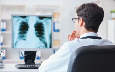
Diagnostic Imaging: Benefits in Cancer Screening
The diagnostic imaging area allows non-invasive observation of the inside of the body through the use of different technologies, tools and specialized medical equipment. This is crucial in the detection of cancer, since facilitates the identification of organ and tissue abnormalities. The main benefits of diagnostic imaging in the early detection of cancer include:
Early detection of tumors before they become clinically apparent
One of the greatest benefits of diagnostic imaging is its ability to detect tumors in early stages, when there are no symptoms or signs yet. that evidence the presence of tumors or irregularities. Thus, by starting treatment early, your success rates are increased.
Accurate assessment and reduction of invasive procedures
Medical imaging provides a more accurate detailed visualization of body organs and tissuesThis helps specialists to differentiate between benign and malignant masses. With this, it is possible to to accurately assess the size, location and characteristics of the tumor. In turn, the need for invasive procedures is reducedas in the case of biopsies.
Monitoring of disease progression and response to treatment.
Diagnostic imaging is not only used to detect cancer, but also to make a monitoring of patients' response to treatment. For this purpose, magnetic resonance imaging or positron emission tomography (PET) tests make it possible to assess whether a tumor is responding correctly to chemotherapy, radiotherapy or immunotherapy treatments. In this way, it is possible to adjust the treatment according to the patient's needs.
Improvement of the patient's quality of life
Imaging studies, being non-invasive techniques, allow for the following detecting cancer without painful procedures or long recovery periods. This improves the patient experience and avoids unnecessary interventions in many cases, improving their quality of life.
Main imaging techniques for cancer detection
There are different medical techniques in the area of diagnostic imaging that play a key role in the detection of different types of cancer:
Mammography
Mammography is the main technique used in the diagnosis and treatment of early detection of breast cancer. By means of a mammography equipment or mammographer, tumors, microcalcifications and suspicious nodules can be identified before they are palpable. We can differentiate two types of tests:
- Screening mammogramsMammography: This is a screening used in women who have no signs or symptoms of breast cancer. Therefore, it is recommended that women over the age of 40 have this type of mammogram as a form of prevention.
- Diagnostic mammogramsBreast cancer screening: It is used when a woman has symptoms such as lumps, pain, discharge or changes in the skin of the breast, or when an abnormality is detected on a screening mammogram or screening mammogram.
Computed Tomography (CT)
Computed tomography, also known as computed tomography, also known as TACis a medical procedure that uses x-rays and digital processing to obtain detailed images of internal organs. It is essential in the screening for lung, liver, pancreatic and colon cancer.
Magnetic Resonance Imaging
In this technique, a magnetic field is used to generate radio waves that allow to create detailed medical imaging of soft tissues. The magnetic resonance imaging is especially useful in the brain, prostate and breast cancer screeningThe results of the tumor assessment are more accurate.
Ultrasound
The ultrasound is a medical procedure that uses ultrasound waves to examine internal organs and structures. This is a key tool in the thyroid, ovarian and prostate cancer screeningIt allows the visualization of abnormal masses without radiation.
Positron Emission Tomography (PET)
The Positron Emission Tomography or PET scanning is a technique that uses a radioactive tracer to identify active cancer cells. It is used in the detection of metastases and in the evaluation of the response to treatment of oncology patients.
Colonoscopy with digital imaging
Allows you to detecting polyps in the colon and rectum that may progress to cancer. The use of colonoscopy in screening programs has significantly reduced colorectal cancer mortality.
The role of technology and Artificial Intelligence in early diagnosis
The advances in technology and the use of the artificial intelligence in medical image analysis are revolutionizing cancer detection. AI-based technologies can analyze mammograms, MRIs and CT scans with high accuracy, making it possible to identify patterns and abnormalities before symptoms become apparent.
The artificial intelligence systems in medicine use advanced algorithms and machine-learning models to analyzing medical images, medical records, genetic data and other sources of information of patients. The use of the AI in medicine improves diagnostic accuracy and efficiency in medical and health care, as large volumes of data can be analyzed quickly and accurately. As a result, it has become a key tool for early disease detection.
Main advantages of AI in cancer diagnosis
- Streamlines the performance of diagnostic imaging studies and interpretation of medical images.
- Offers more detailed and customized analysis for each patient.
- Helps reduce errors.
- Optimizes treatments to suit the needs of each patient.
- Improved health and hospital care.
In the fight against cancer, every small step counts. Prevention, early detection and the use of technology and medical innovation are the most important elements in advancing research into the disease and improving patients' quality of life.
Kiko Ramos
CEO of 4D Médica. Expert in marketing and distribution of medical equipment.
by Kiko Ramos | Feb 5, 2025 | News
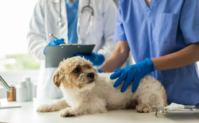
The latest innovations in the veterinary sector have led to improved accuracy and efficiency in the processing of clinical X-ray images in animals. Vieworksa company specializing in medical and industrial imaging solutions, has signed an agreement with the collaboration with SK Telecom Company to accelerate the expansion of integrated veterinary diagnostic solutions.
Through this synergy, Vieworks will be able to integrate its "VXvue" image processing software with the "X Caliber" AI diagnostic assistance service for pets, developed by SK Telecom. The collaboration was signed during the Veterinary Meeting & Expo held in Orlando, USA, on January 15. It involves a expansion of the development of diagnostic assistants based on artificial intelligence (AI)This is a key driver of innovation in pet health care.
What are the uses and functions of the VXvue and X Caliber tools? Below, we analyze each of the tools and the advantages of their integration in the area of veterinary diagnostic imaging.
Vieworks VXvue Software: Advanced Medical Image Processing
The "VXvue" software, developed by Vieworks, is a advanced image processing platform designed to improve the quality and accuracy of medical images obtained from medical imaging detectors. X-rays. Its use is widespread both in human medicine and in the veterinary field, providing optimized tools for analysis and diagnosis.
Key features of VXvue
- High-precision image processingImproves the clarity and detail of clinical images using advanced algorithms.
- Intuitive and easy-to-use interfaceEnables medical and veterinary professionals to analyze images quickly and efficiently.
- Compatibility with different X-ray detectorsOffers flexible integration with digital imaging equipment.
- Customized functions for different types of diagnosticsIt is adapted to the area of human and veterinary radiology, both in the area of small animals and equines.
VXvue applications in veterinary medicine
- Diagnostic imaging in petsProvides detection of musculoskeletal and thoracic diseases in dogs and cats.
- Equine radiologyHigh resolution imaging for bone and soft tissue evaluation in horses.
- Real-time analysisMedical imaging: Provides a detailed study of medical images generated on computers and mobile devices.
X Caliber from SK Telecom: AI-assisted veterinary diagnostics
In 2022, SK Telecom launched "X Caliber", an advanced computer-assisted diagnostic imaging system, with the aim of providing artificial intelligence (AI). It is a tool that allows X-ray images of dogs and cats to be analyzed in about 15 seconds. To do this, it uses cloud technology to store and record data, eliminating the need to install a separate server. Its main objective is to provide veterinarians with a quick and accurate tool for the early detection of diseasesThe quality of diagnosis is improved and veterinary care is optimized.
Features of X Caliber
- Rapid disease detectionIdentifies musculoskeletal, thoracic and abdominal abnormalities in dogs and cats in a matter of seconds.
- AI-based analysis: Uses advanced algorithms trained on large volumes of veterinary data.
- Cloud operationIt is easy and fast to implement, as it works in the cloud. Therefore, it does not require additional infrastructure in healthcare centers and clinics.
- Intuitive and accessible interface: It is compatible with a variety of devices and thus allows for a quick diagnostic review.
- Constant updates and improvementsAnother key aspect is that the use of AI facilitates the expansion of the range of diseases detected.
How does X Caliber work?
The X Caliber tool provides a quick diagnosis in a few simple steps:
- Image captureDigital X-ray detector: A digital X-ray detector is used to capture the medical image of dogs and expenses.
- Analysis with AI in the cloudThe image is sent to the X Caliber platform, where it is processed by artificial intelligence algorithms.
- Anomaly detection in 15 secondsThe system analyzes the image and provides quick results with indications of possible pathologies.
- Viewing on any deviceVeterinarians can review results on computers, tablets or smartphones without the need for additional servers.
Veterinary applications of X Caliber
- Musculoskeletal diseases in dogsDetects bone and joint problems.
- Thoracic pathologies in catsIdentifies pulmonary and cardiac diseases.
- Measurement of heart size (VHS)Allows an accurate analysis of cardiac diseases.
- Detection of abdominal injuriesEvaluates internal organs to identify possible anomalies.
Advantages of VXvue and X Caliber integration
Viewoks' agreement with SK Telecom is aimed at launching a new integrated medical imaging solution that includes X-ray detector, image acquisition software and AI diagnostic support service.. By linking both tools in a single device, it will be possible to analyze clinical X-ray images of dogs and cats, providing abnormal findings for musculoskeletal and thoracic diseases in as little as 15 seconds.
The image post-processingThe use of the information, along with information such as disease location and likelihood of injury, will drastically improve veterinary care. Currently, the diagnostic range of X-Caliber IA is rapidly expanding to include 34 canine and 13 feline pathologies.
Diagnosis of pathologies in dogs and cats
| Species |
Type of Pathology |
Diseases |
| Dogs |
Musculoskeletal diseases (7) |
- Medial patellar dislocation
- Subluxation
- Loss of infra patellar fat pad
- Osteophyte and enthesophyte
- Deviation of the fascial plane
- Fractures
- Enlarged popliteal lymph nodes
|
| Thoracic diseases (10) |
- Generalized cardiomegaly
- Left atrial enlargement
- Diffuse parenchymatous pattern
- Bronchial pattern
- Cranioventral parenchymal pattern
- Caudodorsal parenchymal pattern
- Thoracic mass
- Mediastinal shift
- Tracheal collapse
- Pleural effusion
|
| Abdominal diseases (16) |
- Gastric dilatation
- Gastric Foreign Body
- Dilatation of the small intestine
- Colelites/Hepatolites
- Hepatomegaly
- Microhepatitis
- Splenomegaly
- Kidney stones
- Urinary bladder stones
- Urethral calculi
- Prostatomegaly
- Uterine distension
- Decreased serous detail
- Abdominal mass
- Abdominal wall mass
|
| Measurement of heart size (VHS) |
Yes |
| Cats |
Thoracic diseases (5) |
- Left atrial enlargement
- Parenchymal pattern
- Bronchial pattern
- Cranial mediastinal mass
- Pleural effusion
|
| Abdominal diseases (7) |
- Gastric Foreign Body
- Dilatation of the small intestine
- Hepatomegaly
- Kidney stones
- Urinary bladder stones
- Decreased serous detail
- Peritoneal effusion
|
| Measurement of heart size (VHS) |
Yes |
Through this collaboration, both companies will create synergies and expand sales in the fast-growing global pet healthcare market.
At 4D Médica, we implemented X Caliber AI in the veterinary area.
From 4D MedicalIn the field of veterinary medicine, we are committed to medical innovation, artificial intelligence and technological advances in the field of human and veterinary medicine. In the area of veterinary medicine, one of our latest advances is the extension of the capture panel software for our veterinary direct capture X-ray detectorswhere we will include the X Caliber AI.
This is an AI-enabled extension that includes dynamic updates that can be accessed through membership. In this way, through the use of artificial intelligence, it will be possible to obtain a accurate diagnosis in as little as 15 minutes of a total of 34 pathologies in dogs and 13 pathologies in cats.
In addition to rapid and effective diagnostics, the AI software also will offer personalized recommendations to implement the best treatment depending on the pathology and the diagnosis obtained. This will save both time and resources, optimizing veterinary care to the maximum and improving the quality of life of many pets.
Kiko Ramos
CEO of 4D Médica. Expert in marketing and distribution of medical equipment.

by Kiko Ramos | Jan 3, 2025 | News
Substrate AIa Valencian artificial intelligence company listed on BME Growth, has hired the consulting firm LKS Next to prepare the IPO of its subsidiary 4D Médica.
LKS Next is part of the Mondragon Corporation and has a total of 800 professionals specialized in the industrial, technological and health sectors. Its experience includes mergers, acquisitions and capital markets advisory servicesand, therefore will assist Substrate AI in preparing for the IPO.. From the preparation of the necessary documentation, the choice of the most suitable market for the initial public offering (IPO) and the search for investors, considering the particularities of 4D Médica's business.
Acquisition of 4D Medical by Substrate AI
Substrate AI acquired the 70% from 4D Medical in 2022 for 1.4 million euros. 4D Médica was originally dedicated to the sale of diagnostic imaging hardware for the veterinary sector.under the direction of its founder and CEO, Francesc Ramos, with more than 20 years of experience in the sector.
After the acquisition, the company was transformed into a AI applied to diagnostic imagingwith hardware and software divisions, operating in both veterinary and human medicine.
The purchase of Diagximag is integrated into the subsidiary 4D Médica
In 2023, Substrate AI purchased Diagximaga company focused on hardware for human medicine and main distributor of Samsung in Spain.integrating it with 4D Medical. In addition, we developed a AI-based imaging software to help doctors and veterinarians obtain more accurate diagnoses, which will soon also be available for human medicine.
This software seeks to Improve disease diagnosis and reduce radiation exposure to patients and physicians. by means of collimator self-regulation in ionizing radiation equipment.
4D Médica's sales growth since 2021
Thanks to these initiatives, 4D Médica has tripled its sales in three years1.8 million in 2021 to a figure three times higher in 2024, while maintaining an EBITDA margin of more than 25%.
"We are very satisfied with the road we have traveled in just two years. Together with Substrate AI, and thanks to their technology and support, we have transformed the company and prepared ourselves to become one of the key players in the application of AI to diagnostic imaging, an essential part of current and future medicine," says Francesc Javier Ramos, CEO of 4D Médica.
Therefore, the next step is the joint work of LKS Next, Substrate AI and 4D Médica to ensure that the IPO fits the needs of the company's growth plan.
Kiko Ramos
CEO of 4D Médica. Expert in marketing and distribution of medical equipment.

by 4D Medical | Nov 19, 2024 | News
Thousands of businesses affected, more than 200 deaths and severe damage to many families, businesses and local institutions. The passage of the DANA has had an impact on a total of 75 municipalities in the Valencian Community, two in Castilla-La Mancha and one in Andalusia. Three weeks later, the economic cost is estimated at 1,789 million euros, between repairs and economic reactivation measures, in addition to a further 53 million euros in losses due to inactivity.
According to the data of the evaluation report carried out by the Valencia Chamber of Commerce, the total impact was 1,843 million euros.. But how has it affected the healthcare sector and medical centers?
Retail stores, the main affected by the DANA
The stores and small businesses have been the main affected after the floods. Approximately, a total of 5,228 companies have been directly hit by the DANA, and more than 3,500 businesses have serious damage. For this reason, the government has earmarked a set of measures to repair the damage in the affected premises. The cost of all structural damage will amount to €145 million, in addition to the €394 million earmarked for the cleaning and replacement of assets125 million for the construction of the new plant, such as machinery and furniture, and inventory replenishment.
The healthcare sector and the medical centers concerned
In the healthcare sector, there are many medical, veterinary and dental clinics; physiotherapy centers and health centers which have suffered numerous damages. Floods have affected a multitude of diagnostic imaging and laboratory equipment. and many of them could not be saved after the passage of the DANA.
4D Médica's services to help affected facilities
In view of this situation, from 4D MedicalWe are doing our bit to help all the affected centers. Throughout the last few weeks, we have been carrying out different activities to technical assistance services free of charge. On the one hand, the removal of diagnostic equipment that have had serious mishaps and the reporting on the extent of the damages The companies can process the request for insurance indemnities and also the application for the different state aids.
Main solutions and measures for companies
The retail fabric has been the most affected, especially in the Valencian Community. An impact that could lead to the definitive closure of many businesses, as it is estimated that there is a potential risk that between 25% and 40% of commercial businesses will not reopen. For this reason, the government has promoted a series of economic reactivation measures to support businesses.
Industrial and service establishments have access to a subsidy of up to 7% of the value of the compensable damages36,896 euros. At the same time, it has been approved a 500 million to remove the accumulated sludge debris and repair water networks in the affected municipalities. On the other hand, the victims are exempted from paying the Real Estate Tax (IBI) and have a reduction in the Business Activity Tax (IAE) in 2024.
United to restore clinics and health centers to normalcy
There is still much to be done, but together, we will help restore health and well-being. Many volunteers have volunteered to clean up and bring supplies and materials to the hardest hit areas. At 4D Médica, we are supporting all the companies and centers that need it. The aim is to enable the affected businesses in the healthcare sector to resume their activities.
Together, we will get things back to normal. If your center is one of those affected, you can contact us by the following means:
We will be happy to help you!
by 4D Medical | Nov 5, 2024 | News
The
Principality of Asturias Health Service (Sespa) has received
54 state-of-the-art ultrasound scanners 1.7 million. Following the award in April 2024 of the public procurement contract for the
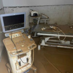
DiagximagOn September 30, the delivery of the equipment was signed. The ultrasound scanners, which were delivered during the months of July and August, represent a very important step forward in the
modernization and digitalization of the healthcare sector in the Asturian region.
How are Diagximag's state-of-the-art ultrasound scanners different?
Diagximag, a subsidiary of Diagximag, an Asturian-based company 4D Medical and belonging to the group Substrate AIhas about state-of-the-art ultrasound scanners which offers a complete imaging solution. These Samsung-branded medical devices combine advanced technology and precision imaging of the different organs, tissues and internal structures of the body. It is an essential tool for diagnosing medical conditions, monitoring the health and development of the fetus during pregnancy, and for guiding certain medical procedures, such as biopsies and tissue extraction. Below, we analyze the main features that define Diagximag ultrasound scanners:
- Equipment includes artificial intelligence and remote controlOne of the innovations of Diagximag ultrasound scanners is that they allow ultrasound scans to be performed using artificial intelligence. They differ in that they include the Sonosync function that allows radiologists to control the equipment completely remotely. In other words, from their own home, they can diagnose patients as if they were present at the medical center.
- High image resolutionThey have a very good image resolution and incorporate Doppler technology, so that tissues and blood flow can be visualized with total clarity. This allows to visualize detailed images and perform a complete diagnosis of the area of the body to be analyzed.
- Intuitive design for multiple uses in the clinical settingIn addition to their multiple functions, ultrasound scanners have an intuitive design that facilitates their use in different clinical environments. A fundamental aspect when it comes to increasing the efficiency of medical diagnostics.
What advantages and innovations do they offer in diagnostic imaging?
The ultrasounds are one of the most widely used medical techniques today, because it is a convenient, inexpensive, safe and non-invasive test. The medical devices used to perform this highly demanded test are ultrasound scanners. They have a rod-shaped toolcalled transducerwhich is responsible for detecting the waves produced inside the body. Through the use of a special gel which is applied to the skin of the area to be examined and the use of a computerThe images are displayed on the screen, which provide the information on fabrics.
The innovative technology based on the application of AI not only enhances the medical diagnostic experience, but also offers a major progress in telemedicine. In this way, the following can be realized rapid diagnostics no matter where the specialist is located. With this, it is possible to reach more regionsas there are many localities that do not have all medical services in health centers. The use of state-of-the-art ultrasound scanners means that more patients can receive a quick and accurate diagnosis, avoiding the need to travel to other regions that have more resources.
The use of these ultrasound scanners incorporating the latest technology provides an improvement in the health sector, so that now the Asturian region will be able to offer an effective and high quality diagnostic imaging, reducing efforts and limitations.