por Luis Daniel Fernádez | Nov 25, 2024 | Equipment analysis
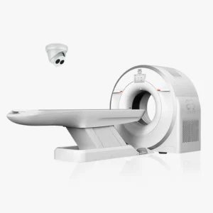
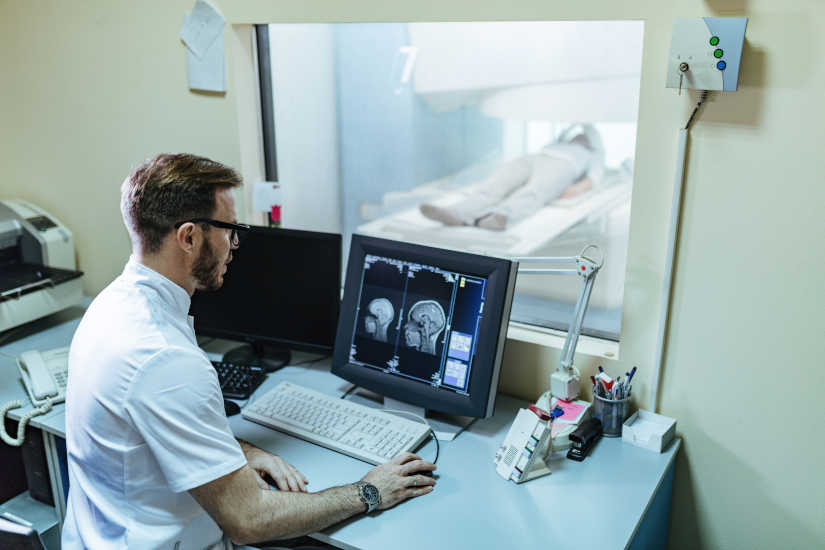
Technology is becoming increasingly important when it comes to storing and managing different data and resources. In the field of medicine, we can highlight the RIS management system for diagnostic imaging. This is a type of specialized software used in the radiology area and in other medical fields to manage information and processes related to the services provided by the image diagnosis. In the following article, we analyze how it works, its main features and advantages.
What is the RIS management system for diagnostic imaging?
The RIS management system is responsible for automating the management of medical imaging data and information. It works like a hospital information system (HIS), but the main difference is that it is specifically tailored to radiology departments in clinics, hospitals and healthcare centers.
It is called RIS (Radiology Information System) and represents a key part of the IT infrastructure in radiology departments, clinics and hospitals. A radiodiagnostic software is a tool that includes a multitude of functions in a single centralized platformfrom manage patient data and history, store medical images and create customized reports. Therefore, it stands out as a solution that helps to improve workflows and optimize medical imaging processes.
Main features and functions of the RIS system
How does the RIS system work? We analyze the main features and functionalities it offers:
Patient registration
Firstly, the RIS system is used to register the patients to be attended. For this purpose, the different data to create your medical record: the personal information of contact, the medical history and the insurance information.
Appointment scheduling
Once the patients are registered in the system, they can be scheduling appointments for diagnostic imaging tests. From radiographs, computed tomography or CAT scans, magnetic resonancesetc. The software organizes and prioritizes orders according to urgency, equipment and personnel availabilityoptimizing the management of time and available resources.
Storage and tracking of medical images
Radiologists can attach the results of the images generated after the medical tests directly to the patient's fileThis speeds up the availability of the studies. At the same time, it also allows include data related to medical examinationssuch as reports and diagnostic information.
Patient follow-up and test management
The RIS system makes it possible to perform the follow-up of the patient's treatment and of the examinations carried out through the system. In this way, the complete medical history can be accessed and patient information can be checked for necessary updates during the diagnostic process.
Workflow tracking
Allows you to track each stage of the process, from the initial request to the generation of the final reportThe system ensures efficient and uninterrupted execution. Another aspect to highlight is that improves collaboration between different medical teams who work in patient treatment, such as radiologists, technicians and medical specialists.
Report generation
Radiologists can writing and sharing diagnostic reports based on processed images. The reports are securely stored and made available to physicians and also to authorized patients. The results are generated digitally, but can also be sent by e-mail and fax, as well as exported for printing on paper. Using the RIS system, different statistical reports can be produced, either for specific examinations, individual patients or groups of patients.
Data analysis and statistics
The system produces reports and statistics on workflows, volumes of studies performed and equipment performanceThe results of this study will facilitate administrative decision making and increase the efficiency of diagnostic imaging services.
Data storage and security
All information, including images, reports, and financial records, is stored in secure databases. This helps to ensure the compliance with medical and privacy regulationssuch as GDPR in Europe or HIPAA in the United States.
Billing and administration
Another of its functions is that automates the creation of invoices related to exams performed. By integrating payment and insurance records, financial management processes can be simplified.
What are the advantages of RIS for diagnostic imaging?
The RIS management system offers numerous advantages, mainly in terms of efficiency, accuracy and quality of service in the field of radiology. We explain its main benefits in the medical field:
Workflow optimization
Allows you to manage all stages of medical diagnosisfrom the request to the delivery of reports. This helps to improve organization and reduce delays that may arise. At the same time, automated appointment scheduling ensures that the efficient use of time and resources.
2. Accuracy and security of data
Reduces the occurrence of errors by centralizing patient information, as test results are located on a single platform. On the other hand, by complying with data security regulations such as HIPAA and GDPR, the medical information included in the RIS system is kept confidential.The patient's data is processed correctly.
3. Quick access to information
Physicians, radiologists and technicians have immediate access to patient records and studiesThis streamlines clinical decision making. And not only that, the system usually includes a integration with cloud-based solutions. In this way, the medical team can remotely access information from anywhere, anytime.
4. Integration with other medical systems
It works in conjunction with other medical systems: both PACS and HIS. On the one hand, the PACS system is used to manage the long-term storage of both images and patient information, and HIS systems are hospital information software used in the management of clinics and hospitals. Therefore, the integration of these systems into the RIS system makes it possible to create a complete healthcare ecosystem.
5. Improved patient care
Offers a agile, comprehensive and seamless patient care experience. Among its advantages is the reduction of waiting times in treatment planning and diagnosis, the results are available more quickly and reduce the administrative burden to be carried out by professionals and patients.
6. Cost reduction
In addition to optimizing the work process, helps reduce costs and increase profitability. It eliminates the need to create paper documentation and reduces administrative errors, thus optimizing billing processes and scheduling of medical services.
Conclusion
The RIS management system is an essential tool for optimizing administrative and clinical processes in radiology and other areas of diagnostic imaging. The use of radiodiagnostic software helps to increase efficiency, service quality and patient care.
Luis Daniel Fernandez Perez
Director of Diagximag. Distributor of medical imaging equipment and solutions.

por 4D Medical | Nov 21, 2024 | Equipment analysis
The X-rays are a form of electromagnetic radiation, similar to visible light. This medical technique was created in 1895 by the physicist Wilhem Conrad Röntgen, whose findings led to the development of radiological practice. It is an essential method in the medical field and is used by means of specific equipment: the X-ray machines. X-rays are able to penetrate matter, so they can pass through most objects and tissues, including the human body. Once through the body, the X-rays reach a radiographic plate or computer where digital images, known as radiographs, are generated.
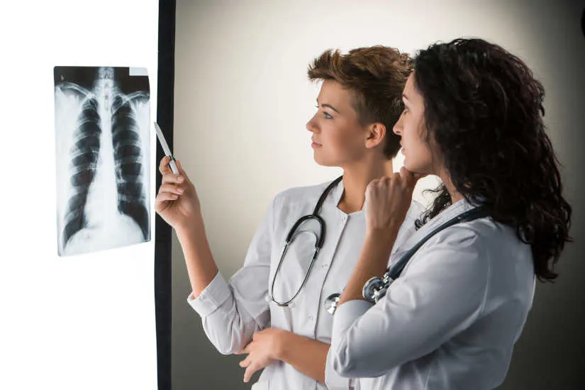
The radiographs are a type of diagnostic imaging and are used to analyze the different internal areas of the organism. The images that are produced are displayed in different shades of black and white.since each tissue allows a certain amount of X-ray beams to pass through it.. Dense materials, such as bones and metals, appear black, while muscles and fatty elements appear in shades of gray. In some types of X-rays, a contrast medium, such as iodine or barium, is introduced so that tissues can be visualized in the images in greater detail.
X-rays can be used aloneas in the equipment of conventional radiology, or combined with other techniquessuch as computed tomography or CT. In the following article, we explain how X-rays work, what they are used for and the types of X-ray machines that exist.
How do X-ray machines and X-rays work?
For imaging in conventional radiography, the patient stands behind a screen that blocks the radiation and operates the X-ray equipment. During the procedure, the body part to be analyzed is placed between the X-ray source and an X-ray detector.
The X-rays passing through the tissues are recorded on a radiation detector plate. Y, depending on the density of the tissues, will pass through a certain amount of radiation.The image produced shows the different degrees of density of the internal structures of the organism. The higher the tissue density, the more X-rays pass through and the whiter the image generated.How are the different tissues displayed?
- Metal has a white color.
- Bone see almost white.
- Fat, muscle and fluids are shown with shadows, in different shades of gray.
- Air and gas are displayed in black color.
Main uses of X-rays
X-rays have multiple uses in the field of medicine. X-rays are used for the diagnosis of diseases and injuries, as a support technique to perform surgical procedures, as a therapeutic treatment, in minimally invasive procedures and for the early detection of diseases. Below, we discuss the different procedures where X-ray technology is used to diagnose and treat diseases:
Diagnostic radiography
X-rays are used as diagnostic test to detect bone fractures, tumors and abnormal masses, pneumonia, etc.as well as lesions, calcifications, foreign objects, intestinal obstructions and dental problems.
2. CT or computed tomography
It combines the X-ray technique together with the computed tomography or CAT scan to create cross-sectional images of the body. Subsequently, can be combined to generate a three dimensional image X-ray images. CT images are more detailed than those of a conventional X-ray and allow professionals to analyze the internal structures of the body from various angles.
Mammography
Breast radiography is used to detecting breast disorders, mainly breast cancer. Breast tissue is sensitive to radiation, so special mammography units are used to minimize radiation exposure. digital radiology equipment.
4. Fluoroscopy
X-rays and a fluorescent screen are used together. to obtain real-time images of the movement inside the body. It is also used to analyze diagnostic processes, such as following the path of a contrast agent.
One of the uses of fluoroscopy is to analyze heart movement and beats. For this, radiographic contrast agents are used to view the blood flow in the heart muscle, blood vessels and organs. This type of technique is also used to guiding an internally threaded catheter during cardiac angioplastya minimally invasive procedure to open clogged arteries that supply blood to the heart.
5. Therapeutic use of radiotherapy for the treatment of cancer.
Another use of X-rays is as therapeutic technique to destroy tumors and cancer cells. The dose of radiation used to treat cancer is higher than the radiation used in diagnostic tests. This type of therapeutic radiation can come from X-ray equipment or radioactive material. that is placed in the body or bloodstream.
Types of X-ray machines
What types of X-ray machines are available on the market? We can differentiate the following medical equipment using this technology:
Conventional X-ray machines
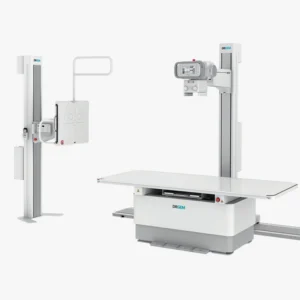
They are the most basic equipment and are designed to obtain static images of the internal structures of the body. It is used for diagnose bone fractures, pulmonary evaluation by means of a chest X-ray and the identification of dental problems.
Portable X-ray machines
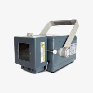
This type of X-ray machines are lightweight, compact and portableThe products can be easily transported. Used in emergencies and rural areasand to care for patients who cannot be transferred.
Digital X-ray machines
They are replacing film plates with digital detectors to develop a real-time diagnostics and the generated images have a high resolution and are of higher quality.
Fluoroscopy systems
These are specific devices that use X-ray technology for the following purposes observing dynamic processes in the body in real time. These machines are used for minimally invasive surgical procedures, gastrointestinal studies and orthopedic diagnostics.
Mammography machines
They are designed for perform breast tissue studies. They are essential for the screening for tumors, abnormalities and breast cancer. In this case, the X-ray emission is low energy in order to better analyze the soft tissues that make up the breasts.
Computed tomography or CT equipment
These units are designed with a advanced system that uses X-rays to create detailed, three-dimensional images of the body. It is highly accurate and is used to evaluate internal injuries, tumors, as well as brain, thoracic, abdominal and extremity studies.
C-arc
These X-ray machines are equipped with a C-shaped arm que emits the X-rays from one end and captures digital images at the other end. C-arc is used for image-guided surgical procedures and in orthopedic and cardiovascular interventions. It offers a deeper analysis, since the area to be analyzed can be visualized from different angles.
Dental X-ray machines
This type of device is designed for capture images of the teeth and various maxillofacial structures. On the one hand, there are the intraoral equipment which capture images of the inside of the mouth and, on the other hand, there are the extraoral equipment which include panoramic systems that take full images of the jaw and mouth. They are mainly used for the diagnosis of caries, periodontal diseases and orthodontic planning.
X-ray machines for bone densitometry
X-rays are used to measuring bone mineral densityand, therefore used to diagnose osteoporosis and perform the follow-up of bone loss treatment.
Meet our 4D Medical equipment
Conclusion
In conclusion, X-rays are a very complete technique that has a large number of uses in the health field and, depending on each medical need, there is specific X-ray equipment to analyze, study and treat various diseases.
If you are interested in purchase an X-ray machine for your clinic, health center or hospital, at 4D Médica we are specialists in the sale of radiological medical equipment and we offer an optimal after-sales service. Ask us about our equipment without obligation.
Contact 4D Médica
Kiko Ramos
CEO of 4D Médica. Expert in marketing and distribution of medical equipment.


por 4D Medical | Nov 19, 2024 | News
Thousands of businesses affected, more than 200 deaths and severe damage to many families, businesses and local institutions. The passage of the DANA has had an impact on a total of 75 municipalities in the Valencian Community, two in Castilla-La Mancha and one in Andalusia. Three weeks later, the economic cost is estimated at 1,789 million euros, between repairs and economic reactivation measures, in addition to a further 53 million euros in losses due to inactivity.
According to the data of the evaluation report carried out by the Valencia Chamber of Commerce, the total impact was 1,843 million euros.. But how has it affected the healthcare sector and medical centers?
Retail stores, the main affected by the DANA
The stores and small businesses have been the main affected after the floods. Approximately, a total of 5,228 companies have been directly hit by the DANA, and more than 3,500 businesses have serious damage. For this reason, the government has earmarked a set of measures to repair the damage in the affected premises. The cost of all structural damage will amount to €145 million, in addition to the €394 million earmarked for the cleaning and replacement of assets125 million for the construction of the new plant, such as machinery and furniture, and inventory replenishment.
The healthcare sector and the medical centers concerned
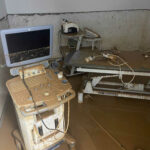
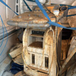
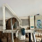
In the healthcare sector, there are many medical, veterinary and dental clinics; physiotherapy centers and health centers which have suffered numerous damages. Floods have affected a multitude of diagnostic imaging and laboratory equipment. and many of them could not be saved after the passage of the DANA.
4D Medica's services to help affected centers
In view of this situation, from 4D MedicaWe are doing our bit to help all the affected centers. Throughout the last few weeks, we have been carrying out different activities to technical assistance services free of charge. On the one hand, the removal of diagnostic equipment that have had serious mishaps and the reporting on the extent of the damages The companies can process the request for insurance indemnities and also the application for the different state aids.
Main solutions and measures for companies
The retail fabric has been the most affected, especially in the Valencian Community. An impact that could lead to the definitive closure of many businesses, as it is estimated that there is a potential risk that between 25% and 40% of commercial businesses will not reopen. For this reason, the government has promoted a series of economic reactivation measures to support businesses.
Industrial and service establishments have access to a subsidy of up to 7% of the value of the compensable damages36,896 euros. At the same time, it has been approved a 500 million to remove the accumulated sludge debris and repair water networks in the affected municipalities. On the other hand, the victims are exempted from paying the Real Estate Tax (IBI) and have a reduction in the Business Activity Tax (IAE) in 2024.
United to restore clinics and health centers to normalcy
There is still much to be done, but together, we will help restore health and well-being. Many volunteers have volunteered to clean up and bring supplies and materials to the hardest hit areas. At 4D Medica, we are supporting all the companies and centers that need it. The aim is to enable the affected businesses in the healthcare sector to resume their activities.
Together, we will get things back to normal. If your center is one of those affected, you can contact us by the following means:
We will be happy to help you!
por 4D Medical | Nov 14, 2024 | Equipment analysis
The computed tomographyalso known as computed axial tomography, also known as computed tomography, or TAChas become one of the most popular techniques of image diagnosis most commonly used. It is a procedure that uses special X-ray equipment and advanced computers to obtain three-dimensional images with different slices of the body.
Since its clinical introduction in 1971, it has undergone successive advances that have allowed its application in different fields of medicine. At present, computed tomography is used to diagnose disorders such as cancer, cardiovascular conditions, infectious processes, trauma and diseases of the locomotor apparatus. In the following article, we analyze how it works, what it is used for and the origin and evolution of this diagnostic test.
How does a CAT scan work?
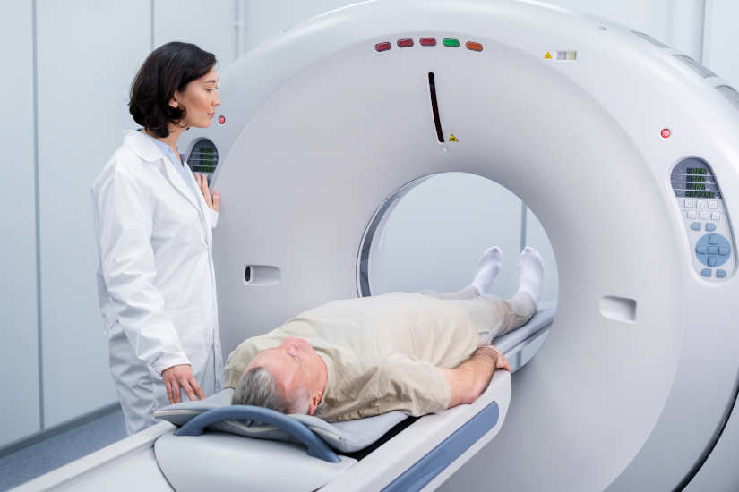
In order to perform this diagnostic imaging, the following is used computed axial tomography system which incorporates a X-ray scanners generating three-dimensional images with different cuts of the interior of the organism.
These slices are called tomographic images and are used for the following purposes study various internal regions of the bodyThe CT scanner can be used to view everything from organs, bones and soft tissues to blood vessels. In contrast to radiography, which only provides a two-dimensional representation, the CT scan makes it possible to observe the three-dimensional images. This makes it possible to analyze tissues with greater detail and clarity. Another aspect to note is that the CT scanner utilizes a X-ray source and has a ionizing radiation higher than that of an X-ray.
During the procedure, the CT scanner rotates around the circular opening of a threaded structure called Gantry. The patient lies on a bed and is inserted inside the scanner so that the specialist can analyze the tissues. The X-ray detectors are located in front of the X-ray source and generate a series of images through different cuts. Subsequently, are transmitted to a computer where the interior of the organism can be visualized and analyzed.
CT contrast medium
As with X-rays, dense structures within the body, such as bones, are easy to image. However, soft tissues are more difficult to image. For this reason, contrast media have been developed that increase the visibility of tissues during X-ray or CT scans. Contain a set of substances that are safe for patients and allow the X-rays to be stopped, so that the organs will be seen in greater detail in the test.
For example, to examine the circulatory system, an iodine-based intravenous contrast medium is injected into the bloodstream to illuminate the blood vessels.
What is CT used for?
CT is used as a clinical diagnostic test, in follow-up studies to analyze the patient's health status, in radiotherapy treatment planning, and even for screening asymptomatic individuals with specific risk factors. A computed tomography scan creates detailed images of the bodywhich include the brain, thorax, spine and abdomen.. Specifically, we can highlight the following uses:
- To help diagnose the presence of a cancer or tumor.. It is one of the most widely used techniques to screen for the presence of colorectal cancer and lung cancer.
- Obtain information about the stage of a cancer.
- Determine if a cancer reacts to treatment.
- To detect the return or recurrence of a tumor.
- Diagnose an infection.
- Support technique to guide a biopsy procedure.
- To guide some local treatmentssuch as cryotherapy, radiofrequency ablation and radioactive seed implantation.
- Radiotherapy planning external beam or surgery.
- Study the blood vessels.
When did computed tomography come into being?
Computed tomography was introduced in 1971 as an X-ray modality. which allowed axial images of the brain to be obtained, so it was a clinical method used specifically in the neuroradiology area. Its evolution has made CT a versatile imaging technique with which three-dimensional images of any anatomical area can be obtained. Currently, it is a diagnostic imaging equipment with a wide range of diagnostic capabilities. wide range of medical applications in oncology, vascular radiology, cardiology, traumatology or interventional radiology.
Evolution: From its beginnings to the present day
At 1971The following were developed first CT scanners for clinical use. During these early years, the EMI-scanner was used, with which brain data could be obtained and the calculation time per image was about 7 minutes in total. Soon after, scanners applicable to any part of the body were developed. At 1973In the early 1990s, the axial scannerswhose equipment had only one single row of X-ray detectors. Subsequently, it was when the helical or spiral scannerswhich incorporated multiple detector rowsits clinical use had a significant impact on the widely used and are the ones currently in use.
Current CT equipment: Main improvements and types
The evolution of the medical equipment has made it possible to obtain significant improvements. In today's systems, the image quality and offer both a better quality of life and a better spatial resolution as a low contrast resolution. In addition, nowadays, the following are also available CT scanners designed for specific clinical applications. Among them, we can highlight:
- Specific CT equipment for radiotherapy treatment planning: These scanners offer a larger aperture diameter than usual, thus allowing a study with a wider field of view. Thus, the images generated have greater detail and clarity.
- Hybrid equipment integrating CT scanners with other imaging techniquesHybrid solutions are now available. Among them, we can highlight the CT scanner incorporating a positron emission tomograph (PET) or a single photon emission tomograph (SPECT).
- Specialized scanners for new indications in diagnostic imagingDual-source" CT scanners, which are equipped with two X-ray tubes, have been developed, as well as "volumetric" CT scanners, which incorporate up to 320 detector rows, making it possible to obtain complete data on the organs analyzed in a single use.
Meet our 4D Medical equipment
Main risks
CT scans can diagnose serious diseases and conditions such as cancer, hemorrhage or blood clots. An early diagnosis is essential in order to find a solution as soon as possible and save lives. However, it is true that it is a test that presents some risks that are important to discuss:
X-Ray
One of the main risks of CT is that it utilizes the X-rayswhich produce ionizing radiation. This type of radiation can have certain effects on the organism and it is a risk that increases with the number of exposures to which a person is subjected. However, the risk of developing cancer by the radiation emitted by the X-rays is generally low.
Use in pregnant women and children
In the case of pregnant women, there are no risks for the baby if the area of the body being imaged is not the abdomen or pelvis. But, medical professionals often perform tests that do not use radiation, such as the magnetic resonance imaging or ultrasound. As for the childrenare more sensitive to ionizing radiationas they have a longer life expectancy and the risk of developing cancer may be higher compared to adults.
Reactions to contrast medium
On the other hand, another aspect to be highlighted is that some patients may have allergic reactions to contrast medium and, in very specific cases, temporary renal insufficiency. In this situation, intravenous contrast media should not be administered to patients with abnormal renal function.
Conclusion
As we have been able to analyze, computed tomography or CT is very useful for detailed and precise analysis of certain internal tissues and organs. By means of X-rays, certain conditions or serious diseases can be studied, which is why it is essential for clinical diagnosis and its application in different fields of medicine.
Are you interested in a CAT scanner? Contact us and we will advise you without obligation so that you can choose the most suitable medical equipment for your clinic or hospital.
Contact 4D
Kiko Ramos
CEO of 4D Médica. Expert in marketing and distribution of medical equipment.


por Kiko Ramos | Nov 12, 2024 | AI in medicine
The use of new technologies and artificial intelligence (AI) has meant a before and after for many sectors. One of them has been medicine, where the latest advances and applications have been influenced by the development of technology. Artificial intelligence is a specialty in the field of computer science that is used to produce programs through a series of algorithms that have the ability to think, learn and make decisions, as humans do.
How does AI work?
AI began to be developed in the 1990s with the aim of creating a computer system that would process data in a similar way to the human brain. One of the branches of artificial intelligence that is most useful in the healthcare sector is the automatic learning. This system has the ability for machines to use the algorithms and learn from the dataThis improves decision making with the processed information.
Through the use of a AI softwareIn addition, a number of functions and tasks can be automated, allowing healthcare professionals to process and analyze medical data more quickly and accurately. This has a significant impact on the different areas of the health care sector and promotes improved healthcare management. Among the main uses offered by AI in the healthcare field, we find that it helps to develop and optimize processes in clinical diagnosis, disease detection and prevention, healthcare, research and the creation or updating of new drugs.
In turn, it has also been a determining factor in the progress of telemedicine and in the development of personalized medical treatments. In the following article, we address the key applications of the AI in medicine and how they are helping to create a more complete, agile and effective healthcare system.
AI applications in medicine
In recent years, AI has been incorporated into medicine to promote higher quality patient care, speed up processes and achieve increased diagnostic accuracy. What are the different areas in which artificial intelligence is currently being used and what improvements have they brought about?
Disease prevention and early diagnosis
AI is a key tool in disease prevention. Through the use of Big Datawhich consists of a combination of digital health data, genomic data and patient behavioral data, can be used as a basis for the development of a new identify risk factors and patterns that lead to the development of certain diseases.
- Spread of diseasesOn the one hand, machine learning algorithms can predict the spread of diseases such as influenza or COVID-19, anticipating epidemic peaks and allowing preventive measures to be taken.
- Detecting signs of chronic diseasesAnother of its applications is that early signs of chronic diseases, such as diabetes or heart disease, can be identified. Chronic diseases are characterized by their slow onset and, in most cases, go unnoticed until they develop into more serious complications. Therefore, the use of AI is very useful for detecting possible signs of disease in medical studies, such as blood tests, ultrasound images or electrocardiograms. In this case, AI algorithms can detect patterns of cardiovascular disease through medical images such as the magnetic resonance imaging or the computed tomography scans.
- Predisposition to genetic diseasesThrough the use of genomic data, artificial intelligence can also analyze predisposition to genetic diseases. AI algorithms are responsible for studying patterns in DNA to identify genetic variants that could indicate a high risk in the development of certain diseases. In oncology, it is used to predict the risk of breast or colon cancer, allowing doctors to design personalized prevention plans.
Clinical diagnosis
In the image processing and interpretation for diagnosisAI offers algorithms that improve the quality and accuracy of clinical diagnostics. They allow to recognize complex patterns in image data automatically, to eliminate noise to increase their quality and to establish three-dimensional models from images of specific patients. In this field, we can highlight the research by IBM researchers on a new study on the use of a new AI model can predict the development of malignant breast cancer.
With rates comparable to those obtained by human radiologists, this algorithm can learn and make decisions about cancer development from imaging data and patient history. Specifically, it was able to predict the 87% of the analyzed cases and was also able to interpret the 77% of noncancerous cases. Therefore, this model could be a fundamental tool to help radiologists confirm or dismiss positive cases of breast cancer.
Personalized medical treatments
Another use of AI in medicine is to find personalized medical treatments for each patient. Based on a set of factors, such as medical history, lifestyle and genetics, the AI algorithms can analyze a large volume of genomic and biomarker data to identify patterns and risk factors.
This can be used to develop a specific medical treatment for the patient's needsThe use of AI in oncology, for example, helps to identify the best treatment for each type of cancer, taking into account the specific genetics of the tumor. For example, in oncology, AI helps to identify the best treatment for each type of cancer, considering the specific genetics of the tumor.
Health care
Patient care is one of the areas where AI can provide great support to both medical professionals and patients. In this case, the AI-based virtual assistants are an ideal solution for automating functions and tasks. These include the appointment management, the realization of basic health consultations, the symptom assessment and the administration of medications.
Promoting telemedicine
In addition, these systems have allowed the evolution of the telemedicine. In this regard, professionals can monitoring patients suffering from chronic diseases remotely and receive alerts of possible anomalies that may arise in their health condition. This offers wide-ranging benefits in reaching a larger number of patients, especially those who live in regions that do not have all the health services in their localities and must travel to receive medical care.
Resource management in medical centers and hospitals
Another area where AI can be implemented is in the management of material and human resources in clinics, hospitals and health centers. Examining large amounts of data from historical records can be essential for to foresee the resources required in a given situationThe company's management and optimization of the available resources can be very helpful for the management and optimization of the available resources. This can be of great help to avoid overcrowding of medical centers at times of high demand and be able to manage the inventory of medical supplies and the availability of beds and medications.
Drug research and development
Artificial intelligence has been fundamental in the development of medical research, both in the development of new drugs as in the optimization of clinical trials. The integration of artificial intelligence into drug design involves a multidisciplinary approach combining both chemistry and biology concepts as well as computer science. to accelerate the discovery of new treatments and medical solutions.
For this purpose, AI models created with machine learning and deep learning algorithms are used to analyze large amounts of data on chemical and biological compounds and the interaction between them.
Robotic surgery
Robotic surgery systems such as the Da Vinci use AI within the field of interventional radiology. In this way, it is possible to perform complex surgical procedures with greater control and precision. These robots are controlled by the surgeons to make small incisions, which helps to reduce the margin of error, perform minimally invasive surgeries and improve patient recovery times..
Another key area in which artificial intelligence can be applied is in the creation of customized surgical plans. In this case, the following are used data from previous surgeries to optimize techniques and to predict possible complications. that may arise during operations.
Training
AI has a key role to play in the technical training of health professionals. It provides multiple tools that help medical specialists to acquire and perfect their skills in different areas, increasing their knowledge in a more efficient and personalized way.
On the one hand, the medical simulations through AI allow students to be able to implementing complex procedures and reducing the risk of errors. At the same time, the following stand out learning platforms that use AI to adjust educational content based on the level of knowledge of the learnerThe aim is to achieve greater efficiency in the learning process.
In summary, AI has a wealth of applications in medicine and there are new improvements and innovations every day that help to further advance the healthcare sector.
Bibliography
APD (n.d.).
Applications of artificial intelligence in medicine. Association for the Advancement of Management. Retrieved from
https://www.apd.es/aplicaciones-inteligencia-artificial-en-medicina/#:~:text=La%20IA%20puede%20acelerar%20el,efectividad%20y%20reduciendo%20efectos%20secundarios.
Sanofi (n.d.). Artificial intelligence in healthcare. Sanofi Campus. Retrieved from https://pro.campus.sanofi/es/actualidad/articulos/inteligencia-artificial-salud
Pakdemirli, E. (2020). Artificial intelligence in radiology: Friend or foe? Radiology, 297(3), 509-510. https://doi.org/10.1148/radiol.2019182622
Sánchez Rosado, E. J., & Díez Parra, A. (2022). Artificial intelligence in medicine: applications and challenges. Industrial Economics, 423, 49-63. Ministry of Industry, Commerce and Tourism. Retrieved from https://www.mintur.gob.es/Publicaciones/Publicacionesperiodicas/EconomiaIndustrial/RevistaEconomiaIndustrial/423/SA%CC%81NCHEZ%20ROSADO%20Y%20DI%CC%81EZ%20PARRA.pdf
International University of Andalusia (2021). Artificial intelligence in medicine: the future of healthcare. UNIA Blog. Retrieved from https://www.unia.es/vida-universitaria/blog/inteligencia-artificial-en-la-medicina-el-futuro-de-la-salud
United States National Library of Medicine (2020). Artificial intelligence in healthcare and the implications for patient safety. JAMA Network Open, 3(4), e200033. Retrieved from https://pmc.ncbi.nlm.nih.gov/articles/PMC7752970/pdf/main.pdf
Mexican Association of the Information Technology Industry (n.d.). Artificial intelligence in healthcare: Digital transformation for healthcare in Mexico. Retrieved from https://amexcomp.mx/media/publicaciones/Libro_IA_Salud_Final_r.pdf
Merly Dayana Jurado-Sánchez, Eddy Maritza Pedroza-Charris, Blanca Mery Rolón-Rodríguez. (2021) How has artificial intelligence helped in medicine. Convictions, 8 (16), 6-20. https://www.fesc.edu.co/Revistas/OJS/index.php/convicciones/article/view/841
Kiko Ramos
CEO of 4D Médica. Expert in marketing and distribution of medical equipment.


