The discovery of X-rays was one of the most important scientific breakthroughs in history. The author of this discovery was the physicist Wilhem Conrad Röntgen, who accidentally discovered this technique in his laboratory in 1895. Over the years, it became a fundamental tool in the fields of medicine, industry and security. The old X-ray machines revolution in the health care sector, particularly in the area of diagnostic imaging. But what is the origin of this medical technique and how did the first X-ray machines come about?
Discovery of X-rays
X-rays were discovered the November 8, 1895 by physicist Wilhelm Conrad Röntgenin Hamburg, Germany. After his studies in medical engineering, he entered the world of physics and obtained his first findings while studying the penetrating power of cathode rays.
Throughout his research, he identified that a nearby fluorescent screen emitted a glow, even though there were solid objects between the radiation source and the screen. This phenomenon indicated that a new form of radiation, invisible to the human eyewas able to pass through opaque objects and project their image on a surface. Röntgen called it "X-rays", using the letter "X" to indicate that it was an unknown phenomenon.
How was the first X-ray created?
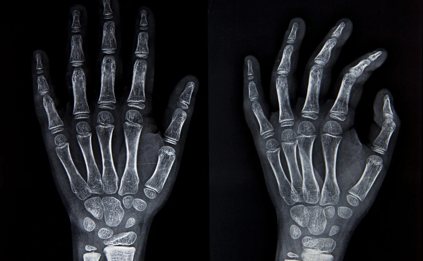
Röntgen, with the assistance of his wife, Anna Bertha Ludwigdiscovered that by holding a lead hoop he could see the bones of his wife's hand. along with her wedding ring. The physical decided to print the image and, to do so, he asked his wife to place her left hand on a metal plate so that he could photograph her, giving rise to the first X-ray.
Findings and initiation of radiological practice
At January 1896, Röntgen published his discovery in the article "On a new kind of rays". A few weeks later, the news spread rapidly around the world and, that same year, the first medical applications began to be developed.
This discovery revolutionized medicine and awarded Röntgen the first Nobel Prize in Physics in 1901.He was the first recipient in the history of these awards. Throughout history, several physicians applied X-ray radiation to treat dermatological conditions and some types of cancer, such as basal cell, uterine cancer and leukemia.
However, the first medical radiologist who did research on its application and the development of radiological practice was Albers-Schönberg. The author produced the first publication on radiology worldwide, entitled "Progress on X-ray areas". Subsequently, the first publications in the field of X-rays began to be developed. X-ray machines.
Antique X-ray machines: Origin, components and characteristics
Currently, X-rays represent one of the most widely used diagnostic imaging technologies. The electromagnetic radiations generated by X-rays have the ability to pass through organic matter and imprint it on a plate with photographic material. Subsequently, they generate medical images in shades of black, gray and white of the internal structures of the human body, giving rise to what is known as the radiography.
The use of this technology allows the diagnosis of multiple diseases and injuriesand, therefore is used in different medical techniques and equipmentBoth in its entirety and in combination with nuclear techniques. From conventional radiography, the computed tomography or CAT scanthe mammographythe fluoroscopy and angiography to bone densimetry.
The first antique X-ray machines were based on the Crookes tube, a vacuum glass device that generated electrons from an electric current. These electrons struck a metallic material, producing X-rays, which could pass through soft tissues and project an image of bones onto a photographic plate.

Components of old X-ray machines
Early X-ray machines were made up of a series of essential components that allowed the generation and capture of images. Unlike modern equipment, the first devices were rudimentary and lacked safety measures, which implied certain risks for both operators and patients.
- Crookes tubeThey worked by means of a vacuum tube with electrodes that generated X-rays upon impact against a metallic material. To do this, they used electrical discharges in low pressure gases.
- High voltage sourceThis element was necessary to accelerate the electrons in the vacuum tube.
- Fluorescent screen or photographic plateIt was in charge of capturing the image projected by the X-rays.
- Manual exposure systemThere was no automatic control of exposure time, which generated a series of risks.
Characteristics of old X-ray machines
In addition to their components, early X-ray machines had several features that set them apart from today's equipment:
- Bulky and fragile structureThey were large and heavy equipment, with glass components that could break easily.
- Prolonged exposure to radiationTo obtain a clear image, patients had to remain still for up to 30 minutes, which increased their exposure to radiation.
- Absence of security measuresLead barriers and protection for operators or patients were not used, since the harmful effects of radiation were not known at that time.
- Low quality imagesThe first radiographs were blurred and with low contrast, which made medical interpretation difficult.
Evolution of X-ray machines
As the risks of radiation were understood, improvements in X-ray technology were introduced:
- 1913 - Coolidge TubeA new, safer and more efficient X-ray tube was developed, allowing better images with less exposure.
- 1920-1930 - Lead protectionLead aprons and protective barriers were implemented to reduce radiation exposure.
- 1970 - Digital radiographyIt allowed obtaining higher quality images with reduced exposure times.
- News - Advanced technologyToday, there are systems such as computed tomography (CT), fluoroscopy and digital mammography, which offer high-precision images with minimal radiation doses.

Risks and limitations of early X-ray machines
Although X-ray machines represented a major breakthrough, they also brought together a set of limitations and dangers:
- High radiation levelsIn the absence of dose control, operators suffered burns and other harmful effects after repeated exposures.
- Burns and diseasesProlonged exposure could cause skin lesions, hair loss and even serious diseases.
- Lack of precisionThe images were of low resolution, which hindered accurate diagnoses.
- Unregulated useIn the early years, there were no regulations on the use of X-rays, which led to accidents and health problems.
Early X-ray machines were a milestone in medical historyHowever, their unregulated use and high level of radiation posed significant risks. Today, X-rays continue to be a major risk. essential tool for medical diagnosis. However, its multiple advances and the use of technology has made it possible to create modern, safe and much more efficient medical equipment for the detection of diseases and other conditions.
If you are interested in acquiring a X-ray equipment or any type of radiological equipment, contact us and we will be happy to advise you and give you the best solution for your clinic or health center.
BIbliography
Faculty of Medicine Gazette UNAM. (2021). For the history of medicine. X-rays. Retrieved February 19, 2025 from https://gaceta.facmed.unam.mx/index.php/2021/07/28/por-la-historia-de-la-medicina-los-rayos-x/
National Institute of Biomedical Imaging and Bioengineering (2022). X-Ray. Retrieved February 19, 2025 from https://www.nibib.nih.gov/espanol/temas-cientificos/rayos-x
Nuclear Forum (2018). History of the first radiography. Retrieved February 19, 2025 from https://www.foronuclear.org/actualidad/a-fondo/historia-de-la-primera-radiografia/
Aulacem. (2022). Infographic: History and evolution of X-Rays. Retrieved February 19, 2025 from https://www.aulacem.es/infografia-historia-y-evolucion-de-los-rayos-x/.

CEO of 4D Médica. Expert in marketing and distribution of medical equipment.
![]()