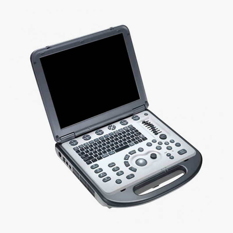
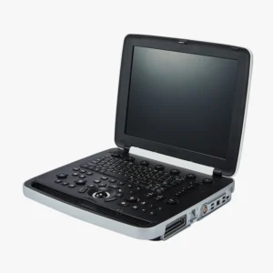
Ecógrafo Mindray M6
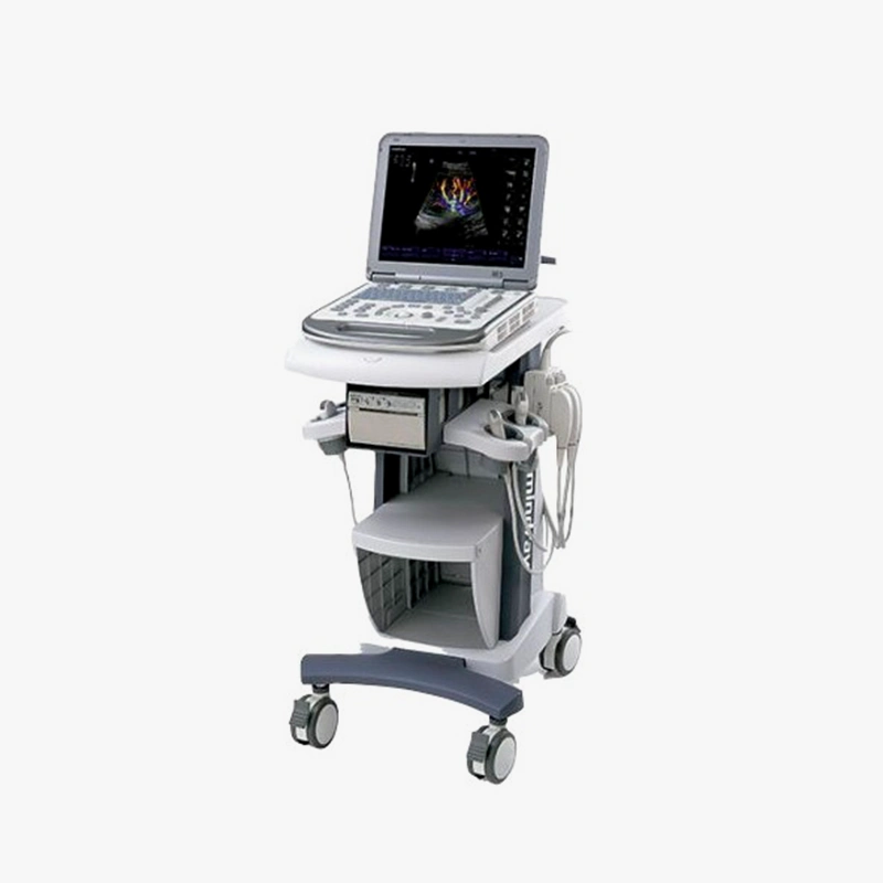
The ecógrafo Mndray M6 es un sistema de ultrasonido portátil diseñado para llevar capacidades diagnósticas de alto nivel directamente a la cabecera del paciente, especialmente en situaciones de cuidados críticos. Mindray ha creado el M6 para ofrecer un equilibrio ideal entre capacidad y tamaño para diagnósticos fiables junto al paciente.
- Advanced visualization with 3D/4D: compatibility with 3D and 4D transducers enables detailed volumetric imaging, crucial for more accurate diagnoses in obstetrics, gynecology and other applications.
- Accurate vascular and cardiac analysis with PW/CW doppler: Pulsed (PW) and continuous (CW) Doppler modes provide detailed blood flow information essential for comprehensive vascular and cardiac assessments.
- Advanced imaging tools: iscape™ and ineedle™:
- Iscape™ (panoramic viewing): the iscape™ function allows the creation of panoramic images of large organs, facilitating the complete visualization of large structures that do not fit in a single image.
- Ineedle™ (biopsy guide): ineedle™ technology improves needle visualization during ultrasound-guided biopsy procedures, increasing the accuracy and safety of the procedure.
- Ideal for demanding environments: its advanced feature set makes it a robust and reliable solution for hospitals, specialized clinics and any medical environment requiring highly accurate and versatile ultrasound diagnostics.
- High quality images: advanced imaging technology for a clear and detailed display.
- Clinical versatility: suitable for various applications such as abdomen, obstetrics/gynecology, cardiology, vascular, small parts, pediatrics and more.
- Optimized workflow: intuitive user interface and efficient examination tools.
- Ergonomic design: facilitates comfortable use over long periods of time.
- Portability (depending on configuration): some models may offer portability options.
- Wide range of transducers: compatible with a variety of transducers for different applications.
- Advanced functions: can include color doppler, power doppler, pulsed doppler, tissue harmonics, spatial composite imaging, etc.
- Display15 inch lcd, high resolution (1024 x 768), led backlight, tilt angle adjustable up to 60°.
- Control panel: alphanumeric keys, function keys, knobs, user-defined keys, 8-segment tgc, trackball (presettable color and speed).
- Transducer ports: 2 or 3 (optional).
- Imaging technologies: ibeam™ (spatial compounding imaging), iclear™ (speckle suppression imaging), pshi™ (phase shift harmonic imaging), tsi (tissue specific imaging), dynamic beam forming.
- Image modes: b, m, color flow mapping (cfm), power doppler imaging (pdi), directional power doppler (dpdi), pulsed wave (pw) doppler, continuous wave (cw) doppler, 3d/4d, free xros m™.
- Data storage500gb built-in hard disk, hard disk image storage, external dvd (optional), istation™ (patient information management system), medsight™, network storage (istorage), cine memory (up to 6197 frames max cine memory).
- Connectivity: 4 usb ports, 1 vga out port, 1 s-video out port, 1 ethernet port (dicom: mpps, print, query/retrieve, store, structured reports, worklist).
- Power supply: 100-240v ac, 50/60 hz, internal power adapter, rechargeable lithium-ion battery (14.8v, 6600 mah, up to 1.5 hours autonomy, charging time < 3 hours).
- Dimensions and weight: 147 mm (depth) x 361 mm (width) x 358 mm (height), net weight: 8.2 ± 0.5 kg (without battery, two probe ports).
This ultrasound scanner is suitable for various medical specialties, including:
- General radiology.
- Obstetrics and gynecology.
- Cardiology.
- Vascular medicine.
- Urology.
- Small parts (thyroid, breast, testicles).
- Pediatrics.
- Internal medicine.
Professional attention
For 4D Médica, product development begins and ends with the customer in mind; it engages clinics, specialty practices and even mobile facilities by working hard every day to deliver high quality patient care.
Maintenance service
The fundamental pillar of the company is a solid and efficient Technical Service team, highly qualified, in continuous specific training in all diagnostic imaging equipment and modalities.
Personalized advice
Implementation studies, implementation reports with calculation of radioprotection barriers, 3D simulation prior to installation and even possible sale. Completely FREE services