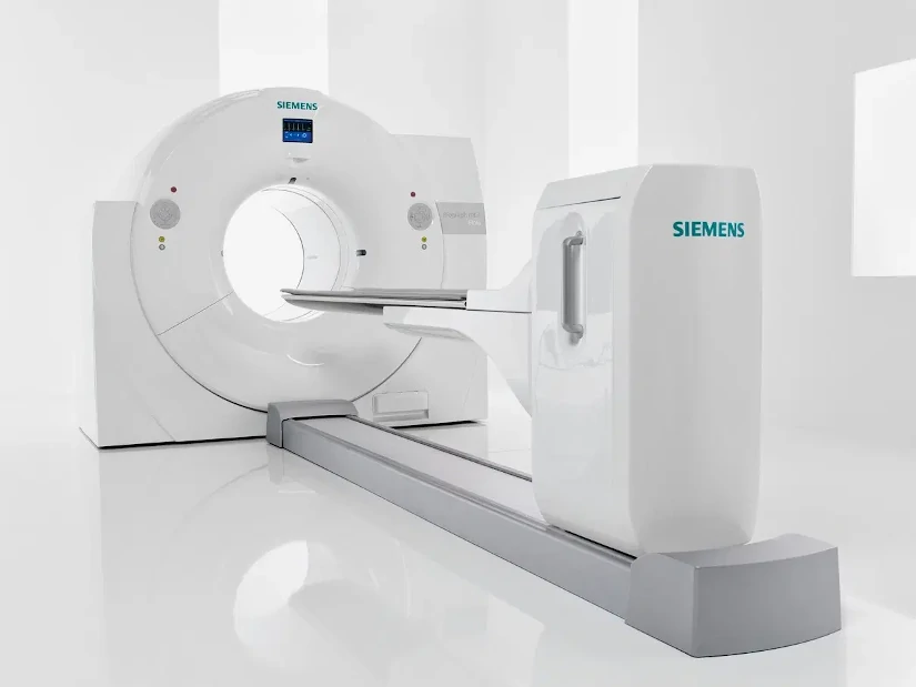
The PET technique CT consists of the integration of two imaging technologies in the same medical equipment: Positron Emission Tomography (PET) and Computed Axial Tomography (CT). The first PET-CT prototype was developed at the University of Pittsburgh in 1998 and its commercialization began in 2001, making it one of the first PET-CT scanners in the world. the most innovative and up-to-date equipment of the area of diagnostic imaging.
A PET CT system is a hybrid medical equipment with a stretcher and a shared medical image acquisition systemThe combination of both technologies provides a tomographic image that represents a cross-section of the organism, offering anatomical and functional information of the interior of the human body. The combination of both technologies provides a tomographic image that represents a cross section of the organism, offering anatomical and functional information of the interior of the human body.
On the one hand, the technology of Positron Emission Tomography or PET scanning provides functional and molecular information of the tissues through the use of a radiopharmaceutical. Therefore, it allows the quantification of various biochemical processes. From cellular metabolism, blood flow and protein synthesis to the analysis of different receptors. For its part, the Computed Axial Tomography or CT reports the different tissue densities generating a high-resolution anatomical image.
Thus, by combining the two techniques in a single integrated PET CT systemcan be generated anatomical and functional images simultaneously. As a result, more complete and efficient clinical diagnoses are offered, both in terms of sensitivity and specificity. Through its ability to detect functional alterations before they are visible in conventional studies, PET CT is fundamental in the early detection of diseases and in the evaluation of the effectiveness of treatments. Especially in enceological, neurological and cardiac diseases. In the following article, we analyze how it works and its main uses in clinical practice.
How does the hybrid PET CT equipment work?
The medical image acquisition protocol in a PET CT study is similar to the procedure for the standard PET technique. In a PET CT scanner, the acquisition of the study consists of three phases: the performance of a topogram, the performance of a CT study that will make it possible to determine the attenuation correction of the PET technique and, finally, the acquisition of the Positron Emission Tomography (PET). Each of these phases is discussed below:
Patient preparation
Before performing a PET CT study, the patient must be properly prepared so that the medical images obtained are of optimum quality. First of all, a radiopharmaceutical is administeredThe most widely used is Fluorine-18-labeled Fluorodeoxyglucose (18F-FDG). This compound makes it possible to detect areas of high metabolic activity that often arise in certain types of cancer, neurological and cardiac diseases. The radiopharmaceutical is administered intravenously and the patient must wait between 45 and 60 minutes for it to distribute correctly by the agency prior to the start of image acquisition.
For optimal uptake of the radiopharmaceutical, the patient must follow a series of medical recommendations:
- Fasting for at least 4-6 hours before the study.which avoids interference with glucose metabolism.
- Staying well hydrated before and after of the procedure.
- Control blood glucose levelsThe high levels may affect the uptake of the radiopharmaceutical.
- Follow instructions from physical rest before the study. Excessive movement prior to the study may generate unwanted FDG accumulation in the muscles.
- In some cases, a controlled breathing protocol to improve the quality of the CT image.
2. Positioning of the patient in the tomograph
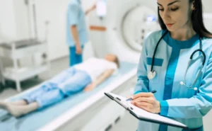
Once the waiting period after injection of the radiopharmaceutical is over, the patient is placed on the bed of the PET CT scanner.. To obtain high quality images and reduce errors in PET and CT image superimposition, it is essential that the patient is well aligned and comfortable. In turn, The patient is asked to extend the arms over the head. if possible, to reduce interference in the images of the thorax and abdomen. On the other hand, metal objects are removed and elements that may affect image quality.
Subsequently, the position of the stretcher is adjusted according to the area to be examined, ensuring that the body is well aligned with the CT scanner detectors. In this process, patient immobility is crucial to avoid blurred images and improve diagnostic accuracy.
3. Making the topogram
The first step in the examination of the patient is to perform a topogram with the PET CT equipment. The images are obtained using the X-rays in a fixed position, which can be anterior, posterior, lateral or in an intermediate orientation. The acquisition is performed with a continuous movement of the stretcher in a predetermined range. In this way, a anatomical image similar to an X-ray projectionwhere the different internal structures and tissues can be analyzed.
It is important that during the procedure the equipment is adjusted and the limits of the PET CT study are defined. Depending on the model of the CT scanner, the fields of view and image formation may be different for different techniques. Therefore, it is necessary to verify that all body parts are within the image with the smallest field of viewwhich are normally those of the CT scan.
4. Elaboration of the TAC study
Once the field of view of the PET CT study has been defined, the patient's stretcher is automatically mobilized to start the CT diagnosis. In the test, a specific breathing protocol is introduced to match the CT and PET image, since the latter is acquired with normal breathing by the patient.
The duration of the CT study depends on various parameters: the extension of the area to be scanned, the rotation speed of the tube and the translation of the stretcher. CT allows detailed anatomical images to be obtained through the use of X-rays, which facilitates the precise localization of organs and structures. In some cases, a contrast medium may be administered to enhance the visualization of vascular structures or specific lesions.
Regarding its duration, a full body CT study using the hybrid equipment is less than one minute. This is because the images obtained are used for attenuation correction in the PET study, which significantly reduces the acquisition time. In PET equipment, when germanium (Ge) sources are used, the CT procedure time amounts to 20 to 30 minutes. With this, radiation exposure is reduced and the patient experience is improved.
5. Acquisition of the PET study
After the CT analysis, PET images are acquired, in which the metabolic data of the tissues are captured. For this purpose, the couch is moved to position the patient in the field of view of the PET scanner, encompassing different positions on the stretcher to cover the region of interest to be analyzed. All these areas are the ones that cover the range explored by CT.
The acquisition time of the PET study can range from between 10 and 30 minutes. This depends on the stretcher positions, the range scanned, as well as the equipment used. During this phase, the areas of the body with abnormal metabolic activity are highlighted on the PET imageThis makes it possible to detect tumors, infections or neurological problems with great precision.
6. PET CT image reconstruction
Reconstruction is performed in parallel to image acquisition.This allows results to be obtained in just a few minutes. This step is essential to generate highly accurate fused images, combining the metabolic information from PET with the detailed anatomical structure from CT.
In this process, the reconstruction time of each CT slice is less than one second.The PET images are reconstructed and available for analysis at the end of the acquisition of the last couch position. To achieve this, we use the reconstruction algorithms available in PET tomographs with the scatter and attenuation corrections determined from CT images.
7. Image analysis and interpretation
Once the images have been reconstructed, they are analyzed, where specialists can analyze different types of medical images:
- PET images without correctionThey show the uptake of the radiopharmaceutical in the body.
- Corrected PET imagesThey incorporate attenuation adjustments to improve accuracy.
- CT imagesThey offer anatomical details of the explored region.
The image fusion software allows the superimposition of PET and CT information, facilitating the exact localization of lesions and their subsequent analysis and interpretation.
What is PET CT used for?
It is a diagnostic technique that is essential in different medical specialties:
- OncologyEarly detection of tumors, evaluation of metastases and treatment follow-up.
- NeurologyIt is used for the diagnosis of diseases such as Alzheimer's, Parkinson's and epilepsy.
- CardiologyThey play an essential role in the evaluation of blood flow and the detection of lesions and abnormalities in the heart.
- Immunology and infectionsHelps in the identification of inflammatory processes and infectious diseases.
Source || Canva
Clinical applications of PET CT
PET CT technology combines the advantages of an anatomical and a functional imaging technique. In the current medical context, the use of this hybrid equipment is used in the following cases:
To confirm or rule out a malignant tumor pathology.
The PET technique can to analyze whether a lesion is benign or malignantThis can avoid the need for biopsies and other invasive diagnostic tests. In turn, it allows early detection of tumor processes, before anatomical changes occur that can be detected by morphological imaging techniques.
Determine tumor extent
It has the ability to perform whole body studieswhich makes it possible to rule out or confirm other malignant lesions concurrent with the primary tumor.
Detecting new tumor recurrences
Through this technique, it is possible to differentiate between malignant processes and new tumors that arise recurrently. This can be used to optimize patient treatment planning.
Assess response to treatment
The metabolic changes produced before an adequate response to chemotherapy are observed earlier in PET imaging than in other techniques. diagnostic imaging. Therefore, this type of medical imaging is an early indicator of tumor response. Their use helps to determine the continuation of certain treatments or, on the contrary, their interruption.
Conclusion
The use of hybrid PET-CT equipment is a crucial advance in medical diagnostics. It combines a functional and anatomical analysis of the inside of the human body in a single medical device, making it fundamental in the early diagnosis of cancer and other neurological and cardiological diseases. The combination of technology and medicine continues to save lives, and the PET CT technique is a clear example of this.
If you want to get information about PET-CT or other radiodiagnostic equipment, you can contact us. Our 4D team will give you advice to find the best solution for your clinic or hospital.
Bibliography
Medigraphic (2005). Acta Médica Article. Retrieved February 13, 2025, from https://www.medigraphic.com/pdfs/actmed/am-2005/am053e.pdf
Electronic Journal of University Medical Science (n.d.). Article in RECIAMUC. Retrieved February 13, 2025, from https://www.reciamuc.com/index.php/RECIAMUC/article/view/1326/2074
Medigraphic (2014). Acta Médica Article. Retrieved February 13, 2025, from https://www.medigraphic.com/pdfs/actamedica/acm-2014/acm141i.pdf
Martínez del Valle, M. (2016). PET-CT: Physical basis, instrumentation and technological advances. Radiology, 58(5), 377-389. Elsevier. Retrieved from https://www.elsevier.es/es-revista-radiologia-119-articulo-pet-tac-bases-fisicas-instrumentacion-avances-S0033833816301801?newsletter=true
Spanish Society of Nuclear Medicine and Molecular Imaging (SEMNIM). (2019). PET-CT protocol. SEMNIM. Retrieved February 13, 2025, from https://www.semnim.es/wp-content/uploads/2019/07/79.pdf

Director of Diagximag. Distributor of medical imaging equipment and solutions.
![]()