The mammography is a technique of diagnostic imaging which uses a system of low-dose X-rays to examine the inside of the breasts. This is a medical test that consists of performing a breast radiography. When performing a mammogram, a mammography machine is used. specific equipment: the mammograph.
It is a medical equipment that is specifically designed to capture X-ray images with a high resolution to detect signs and irregularities in breast tissue. The design and the different parts of a mammography equipment allow a minimum dose of radiation to be used during the test, making it an effective, fast and safe examination.
Health professionals use this test to look for early signs of disease in breast tissue. Among them, breast cancer. The mammography test is called mammogram and its main purpose is to detect abnormalities such as tumors, cysts or microcalcifications in the breast. We analyze, below, what mammography consists of, how the mammogram works and its different parts.
Mammography: What is mammography and types of mammograms?
The use of the mammograph is used as a screening tool for early detection of breast cancer in womenA mammogram is used both in women who have no symptoms and to diagnose the presence of abnormalities in women who notice breast irregularities. A mammography examination or mammogram exposes the woman to a small dose of ionizing radiation to generate medical images of the inside of the breasts. We can differentiate between two types of mammography:
Screening mammography
A screening mammogram is performed in women who have no signs or symptoms of breast cancer. This type of mammography should be performed periodically in women from the age of 40 as a form of prevention. By means of this diagnostic test, it is possible to detect irregularities in the breast tissue, such as tumors, cysts or microcalcifications. Screening for breast disease at early stages, especially breast cancer, provides a range of advantages:
- Allows the identification of tumors before they are palpable. or present visible symptoms.
- Enables treatment to be initiated in the early stagesbefore the disease has spread.
According to different studies, it has been proven that the screening mammography screening decreases breast cancer morbidity rates by detecting the disease at treatable stages, increasing the chances of successful treatment.
2. Diagnostic mammography
Diagnostic mammography is used when a woman presents symptomsas lumps, pain, discharge or changes in the skin of the breast. It is also used when an abnormality is detected on a screening mammogram or detection. This type of examination allows the affected area to be studied in greater detail and thus identify whether the breast condition is benign or malignant.

Mammograph operation
The medical equipment The mammogram is a specialized medical device that allows the analysis of breast tissue and the presence of abnormalities. This is specialized medical equipment that uses X-rays to generate medical images of the inside of the breasts. How a mammogram works consists of several stages:
Preparation of the patient
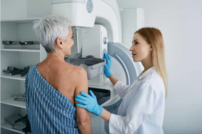
The process begins with the positioning of the patient in front of the mammograph. During the mammogram, a radiology professional will positions the breast on a flat platform of the mammography equipmentwhere the breast will be gradually compressed. The specialized technician will guide the patient to ensure proper posture and perform the medical test.
2. Breast compression
Once the breast is positioned, an adjustable compressor descends to press on the breast tissue gently, but firmly.
3. X-ray emission
The tube of X-rays of the mammogram emits a controlled beam of radiation passing through compressed breast tissue. This radiation is absorbed to a greater or lesser extent depending on the density of the tissue:
- The dense tissuessuch as tumors or microcalcifications, absorb more radiation. They appear clearer and brighter in the images.
- On the other hand, the fatty tissues absorb less radiation and appear darker.
4. Image capture
The radiation passing through the breast is captured by a detector which transforms the data into a digital image or radiographic film. Modern mammographs are often equipped with digital technology that allows images to be stored and processed on a computer.
Subsequently, these generated medical images can be integrated in the RIS system to automate the management of medical imaging data and information, facilitating its analysis and comparison with previous studies.
5. Variation of angles and views
To ensure a complete evaluation of the breast tissue, images are captured from different angles. The different perspectives help physicians identify abnormalities that may not be visible in a single view. The views that are analyzed in a mammography study are:
- Craniocaudal (CC)This is a top-down view.
- Mediolateral oblique (MLO)This type of slanted view allows a greater amount of breast tissue to be studied, especially that close to the axilla.
6. Image analysis
Once the images have been obtained, a specialized radiologist reviews the results for possible abnormalitiesas cysts, calcifications, tumors or suspicious tissue changes. Nowadays, digital images offer many advantages, since they allow adjusting contrast and brightness to improve image quality, obtaining a more efficient and accurate diagnosis.
The mammograph: Parts and components
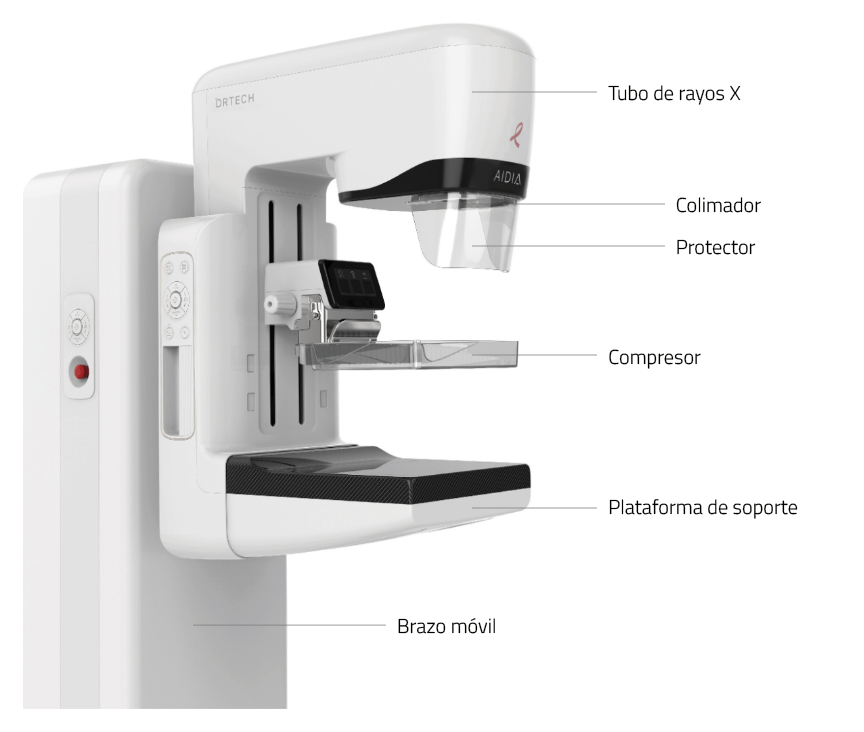
A mammogram is composed of several elements that work together to ensure clear and accurate images. Each component has a specific function that contributes to the quality of the diagnosis and the safety of the procedure. What are the main parts of a mammography machine?
X-ray tube
The X-ray tube is the component responsible for generating the X-ray beam that passes through the breast tissue. and subsequently produce high quality images. The mammograph uses a lower radiation doses than standard X-rays. This is because, since x-rays do not pass through this area easily, the mammography equipment is designed with two plates that compress and flatten the breast to separate the breast tissue. In this way, a higher quality medical image can be created and the amount of radiation during the exam can be reduced.
2. Compressor
The compressor is a movable plate that descends to press the breast against the mammography platform. Its function is to compress the breast tissue gently and firmly, providing the following advantages:
- Reducing the thickness of breast tissue to improve the visualization of internal structures.
- Minimizing X-ray scatteringThe image quality is improved.
- Avoid blurred images caused by the involuntary movement of the patient.
- Allowing the use of a lower dose of radiationmaking the procedure safer.
3. Support platform
The support platform is a flat surface on which the breast is positioned during mammography. It provides a stable and firm foothold, ensuring that the breast tissue is correctly positioned for sharp, detailed images.
4. Detector
The detector is the component that captures the radiation passing through the breast tissue and converts it into an image.. Depending on the type of mammograph, it can be of different types:
- DigitalX-ray: Converts X-rays into electronic data that is processed and stored in a computer, facilitating detailed and rapid analysis.
- Radiographic filmThis type of detector is used in analog mammographs, where the image is printed on a special film.
5. Collimator
The collimator is a structure that directs and confines the X-ray beam to the specific area of the breast that needs to be examined. This component prevents other areas of the body from receiving unnecessary radiation, making the procedure safer.
6. High voltage generator
The high-voltage generator is responsible for supplying the energy necessary for the X-ray tube to function correctly. It regulates the intensity and duration of the X-rays, adapting to the needs of each scan.
7. Control station
The control station is the panel or computer from which the technician operates the mammography machine. Allows you to adjust the parameters of the examinationIt also ensures that the procedure is performed in a precise and customized manner for each patient. It also ensures that the procedure is performed accurately and customized for each patient.
8. Positioning system
The positioning system includes mechanisms for adjusting the height, tilt and angle of the mammography machineThe system can be adapted to the physical characteristics of each patient. This system facilitates the imaging from different perspectivesobtaining a complete analysis of the breast tissue.
9. Image processing software
In digital mammographs, the digital mammogram processing software medical images is an advanced tool that improves the quality of captured images. Allows adjustment of contrast, brightness and other parameters to highlight specific details, as well as compare current images with previous studies, facilitating a more accurate diagnosis.
10. Security system
The mammogram is equipped with a safety system that ensures that radiation exposure is minimized and safe for the patient. In addition, some devices are equipped with sensors that automatically stop the process if a problem is detected technical or positioning.
Advantages of mammography
The mammograph is an essential medical device for the detection, diagnosis and follow-up of breast diseases, especially breast cancer. Its use not only allows early identification of abnormalities, but also contributes to more effective treatment planning. What are its main advantages?
Prevention and early detection of diseases
The mammograph is capable of identify abnormalities in breast tissue in early stages or even before symptoms and signs are visible. The early detection is key to significantly increasing the chances of successful treatment, as it allows the disease to be addressed before it develops to an advanced stage.
In turn, the periodic mammograms are performed is a fundamental strategy for the prevention of breast cancer in women. By detecting breast cancer in its early stages, it helps to reduce the mortality associated with this disease and improves the quality of life of patients.
Non-invasive, fast and safe procedure
Mammography is a non-invasive diagnostic procedure that uses a minimal dose of X-rays, meeting strict safety standards. The mammography exam is fast and efficient. It usually has a duration between 10 and 30 minutesdepending on the type of mammography performed:
- The screening mammogramsDuration: Its duration is between 10 and 20 minutes.
- The diagnostic mammogramsThey have a longer life, between 15 and 30 minutesThey include different views and images to analyze the area in a specific way.
High precision imaging
Modern mammographs, especially digital mammographs and those using 3D technology (tomosynthesis), provide high-resolution images that allow the breast tissue to be analyzed in great detail. This precision facilitates the detection of small or subtle irregularities and improves the differentiation between normal tissues and abnormalitiesreducing the probability of false positives or negatives.
Examination customization
The design of the mammograph allows tailoring the procedure to the individual characteristics of each patient. Exposure parameters, X-ray intensity, acquisition angle and compression level can all be adjusted. All this allows you to generate high quality medical images and optimize the patient experience.
Fast and efficient diagnostics
The mammogram streamlines the diagnostic process by generate medical images in a short period of time. In this way, when abnormalities are detected, physicians can immediately plan further studies and start treatment as soon as possible.
Multiple uses and clinical applications
In addition to being a key tool for the early detection of breast cancer, the mammogram has also other important applications:
- Monitoring of the evolution of oncological treatments.
- Performing image-guided biopsiesThis improves the accuracy of the procedure.
- Identification of benign changes or non-malignant disease in the breast tissue.
Conclusion
The mammograph is an advanced technological tool that combines precision, safety and efficiency for the detection and diagnosis of breast diseases.
If you need advice on radiodiagnostic medical equipment, at 4D we help you choose the most appropriate solution that suits your clinic's needs and budget. We have new and second hand equipment, as well as renting and leasing options. Contact us without obligation.
Bibliography
RadiologyInfo.org (n.d.). Mammography. Retrieved January 15, 2025, from https://www.radiologyinfo.org/es/info/mammo
MedlinePlus (n.d.). Mammography. U.S. National Library of Medicine Retrieved January 15, 2025, from. https://medlineplus.gov/spanish/mammography.html
Centers for Disease Control and Prevention (CDC). (n.d.). Mammograms. Retrieved January 15, 2025, from https://www.cdc.gov/breast-cancer/es/about/mammograms.html
Revista Argentina de Mastología (2020). Importance of mammography in the early detection of breast cancer. Retrieved January 15, 2025, from https://www.revistasamas.org.ar/revistas/2020_v39_n141/06.pdf


