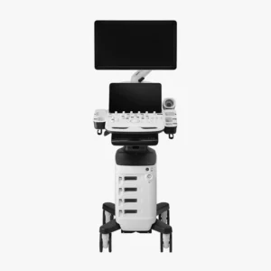
At present, the ultrasound scanner has established itself as a fundamental tool in the area of diagnostic imaging. This device uses the ultrasound technology to obtain accurate, real-time images of the internal structures of the human body, facilitating the evaluation of organs, tissues and blood vessels. without the need for invasive procedures.
The ability of ultrasound to provide detailed, safe and rapid information has revolutionized clinical practice. The use of this medical equipment enables healthcare professionals to detect and monitor a wide variety of pathologies in an early and effective manner. In addition, its versatility and portability has extended its use to multiple medical specialties.
The use of the ultrasound scanner is used to perform ultrasounds in order to analyze organs and tissues internally. It is one of the most widely used medical techniques, as it is a fast, effective and non-invasive method. It is mainly used to detect diseases and anomalies, to monitor the health of patients, to study the development and growth of the baby during pregnancy, as well as to guide certain medical procedures.
In contrast to other imaging techniques, such as X-rays or the Computed Axial Tomography (CT) scanultrasound does not use ionizing radiation, which makes it a safer technique. In addition, its portability and ease of use has enabled its integration in consultation rooms, emergency rooms and intensive care units, facilitating real-time clinical decision making and improving patient care.
In the following article, we analyze the origin of this medical equipment up to the present day, how an ultrasound scanner works, as well as its applications in clinical practice.
Origin of the ultrasound scanner: From its beginnings to the present day.
The development of the ultrasound scanner is closely linked to the development of ultrasound technology and its application in the medical field.

Early studies: Discovery of the piezoelectric effect
The first studies on ultrasonic waves date back to the end of the 19th century, when the French physicists Pierre and Jacques Curie discovered in 1880 the piezoelectric effect. This physical phenomenon consists of the ability of certain materials, such as quartz and some ceramic crystals, to generate an electrical charge when subjected to mechanical pressure.
The importance of the piezoelectric effect in ultrasound is fundamental, since it constitutes the basis of ultrasound transducer or probe operation. In practice, piezoelectric crystals located in the transducer convert electrical signals into ultrasonic vibrations (ultrasound waves), which are transmitted to the patient's body. Through the piezoelectric effect, these echoes are transformed into electrical signals that are processed by the ultrasound scanner to generate images in real time.
Development of the first ultrasound scanner: From the piezoelectric effect and ultrasound to the medical field
Following the discovery of the piezoelectric effect, the phenomenon was initially applied in industrial and military fields, particularly in the development of sonar devices for underwater detection during World War I and World War II.
However, the potential of ultrasound and the piezoelectric effect to generate and receive acoustic waves did not go unnoticed by the scientific and medical community. The adaptation of this technology to the medical field began in the middle of the 20th century. The Scottish physician Ian Donald, together with engineer Tom Brownwere the pioneering researchers who began to apply the piezoelectric effect to clinical explorationThe first of its kind, it laid the foundations of medical ultrasound.
Specifically, it was in the 1950s, when researchers developed the first clinical ultrasound prototype. Initially, ultrasound was used in obstetrics to visualize the fetus and detect pathologies during pregnancy, which was a revolution in prenatal monitoring.
From the 1960s and 1970s to the present day: Advances in ultrasonography
During the 1960s and 1970s, ultrasound technology made significant advances. It became from still images to real-time ultrasound scanswhich made it possible to observe the movement of internal organs and structures. Subsequently, the incorporation of the Doppler effect made possible the study of blood flow and vascular evaluation.The new ultrasound system has been designed to further expand the clinical applications of the ultrasound scanner.
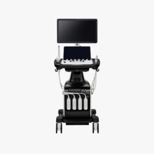
In recent decades, the development of technology and information technology has made possible the emergence of more compact, portable ultrasound scanners with higher image resolution. Today, the ultrasound scanner is a safe, effective and versatile tool used in a wide variety of specialties, from emergency medicine to cardiology, gynecology and internal medicine. As a result, it has become an indispensable piece of medical equipment in medical practice.
Next-generation ultrasound scanners: innovation, technology and artificial intelligence
In recent years, technology has advanced greatly in the field of medicine. The state-of-the-art ultrasound scanners offer images in 3D, 4D and 5D technologyand therefore allow for visualize the inside of the human body in motion and in real time.
One of the most recent innovations is the ultrasound scanners that incorporate digital processing systems that apply the artificial intelligence in medical image analysis. The use of a AI software in ultrasound equipment provides greater speed, efficiency, safety and diagnostic accuracy, providing advanced and detailed analysis that improves clinical decision making.
In this area, it is worth mentioning the use of state-of-the-art ultrasound scanners to visualize the fetus in real time. It is known as emotional ultrasound and allows parents to get to know the baby before it is born. This type of ultrasound combines the 3D technologywhich provides three-dimensional images, with 4D and 5D technologyThe newborn's movements can be seen in real time with high image clarity and quality. With this, the baby's main movements can be seen. From yawning, opening the eyes and moving to changing position.
How an ultrasound scanner works: Step-by-step procedure
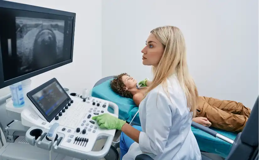
The ultrasound scanners are an essential tool in medical practice. Understand its operation and the workflow during an ultrasound examination is essential to ensure diagnostic quality and patient safety. Below, we discuss how an ultrasound scanner works and the step-by-step procedure:

1. Preparation of the patient and application of the conductive gel
First of all, the patient is instructed on the position The position to be used depends on the area to be scanned and the type of diagnosis to be made. Before starting the scan, the conductive gel on the patient's skin. It has the function of eliminating the air that is generated between the skin of the area to be examined and the transducer or ultrasound probe, facilitating the transmission of ultrasonic waves.
Selection of transducer type
One of the most important components of the ultrasound scanner is the transducer or probe. There are different types of transducersEach is designed to scan different regions and depths. While linear transducers are used for vascular and superficial studies, convex models are useful for deep abdominal scans.
Therefore, the medical professional will be in charge of selecting the type of transducer, connect it to the equipment and verify its correct operation. before starting the study.
3. Ultrasound emission and reception
Once the transducer is prepared, the operator places it on the gel-covered area. The transducer emits high-frequency ultrasound waves that penetrate the patient's internal tissues. When these waves pass through the body and are reflected at the interfaces of the different tissues and organs, the reflected waves, known as echoes, return to the transducer..
4. Echo pickup
The transducer also acts as a receiverby detecting the reflected waves (echoes) that are generated from the various internal structures. These echoes contain information on the location and characteristics of the traversed tissuesThis allows us to analyze the state and functioning of the different organs.
5. Image processing
Echoes picked up by the transducer are converted into electrical signals.These images are processed by the ultrasound console through different algorithms. The result is the generation of two-dimensional or three-dimensional images in real time that are displayed on the screen of the equipment.
By analyzing medical images, the operator is able to observe the anatomy and movement of internal organs and structures. In turn, with the use of the Doppler mode, the blood flow in the tissues can be studied.
6. Systematic exploration
The professional performs a methodical sweep by moving the transducer over the area of interest to be analyzedThe different sections (longitudinal, transversal, oblique) are obtained to fully examine the organs and structures. This systematic exploration is the key to obtain a complete and detailed diagnosisThe results of the study should be documented in a way that does not omit relevant findings, and the results should be adequately documented.
7. Adjustment of image parameters
During scanning, the operator can adjust various parametersThe display mode (2D, 3D, Doppler) can be set from the gain (brightness), depth and focus to the display mode (2D, 3D, Doppler). In this way, you can optimize image quality and adapt it to the anatomical characteristics of the patient.
8. Interpretation of medical images
After performing the examination, the physician is in charge of analyze the images obtained in real timeThe data can be used to identify possible alterations and to make static captures or recordings of relevant sequences. By means of these recordings, the final report can be fully documented, which will serve as the basis for the diagnosis and the clinical decision making.
9. Completion and cleaning
At the conclusion of the study, the operator removes the gel from the patient's skin. Subsequently, a protocol for disinfection and cleaning of the equipment usedThe transducer and the contact surface between each patient.
This structured process makes ultrasound a fast, safe, non-invasive and highly versatile technique, facilitating the accurate assessment of multiple organs and pathologies in daily clinical practice.
Main clinical applications of ultrasound
The use of ultrasound scanners covers practically all medical specialties. The main clinical applications include the following areas:
Obstetrics and gynecology
Ultrasound is essential in the pregnancy monitoringIt is used to evaluate fetal development, the location and viability of the pregnancy, the detection of congenital anomalies and the control of obstetric complications. It is also used for the study of gynecological pathologiesas ovarian cysts, uterine myomas or endometrial alterations.
Cardiology
The echocardiogram is an essential technique for the assessment of cardiac anatomy and functionallowing to diagnose valvular diseases, cardiomyopathies, heart failure, congenital heart diseases and evaluate blood flow using the Doppler mode.
Internal medicine and gastroenterology
The abdominal ultrasound allows examination of organs such as the liver, gallbladder, pancreas, kidneys, spleen and bladder. It therefore plays a key role in the diagnosis of masses, cysts, stones, inflammations and other pathologies. It is also used in the assessment of ascites and in the control of interventional procedures.
Vascular exploration
Through the Doppler ultrasoundthe blood flow in arteries and veinsIt is therefore used in the diagnosis of deep vein thrombosis, venous insufficiency, arterial stenosis, aneurysms and other vascular diseases.
Musculoskeletal
Allows you to study muscles, tendons, ligaments, joints and soft parts, facilitating the diagnosis of sports injuries, tears, tendinitis, bursitis, hemorrhages and subcutaneous masses.
Urology
It is used for assess the prostate, bladder, testicles and kidneysbeing useful in the diagnosis of prostatic hyperplasia, lithiasis, tumors and other urological alterations.
Pediatrics
Ultrasonography is especially useful in the study of pediatric pathologies, such as the hip dysplasia, hydrocephalus, renal malformations and abdominal alterations in newborns and infants.
Guidance on interventional procedures
The ultrasound scanner facilitates the performance of biopsies, drainages, punctures, catheter placement and other interventionsThe procedure is safer and more accurate.
Emergency medicine and intensive care
Its speed and portability allow immediate diagnosis of serious pathologies such as pleural effusions, hemoperitoneum, pneumothorax, cardiac tamponade and rapid assessment in polytraumatized patients (FAST ultrasound).
Conclusion
Ultrasound has established itself as an essential tool in clinical practice.It offers an accurate, efficient, safe and real-time medical analysis of the different internal organs and tissues. Its non-invasivenessThe absence of ionizing radiation and its versatility to be adapted to multiple specialties make it an indispensable resource both in the initial evaluation and in the follow-up of numerous pathologies.
Your portability and speed facilitate clinical decision making in a variety of settings, from outpatient to emergency situations. From its origins to the present day, the use of ultrasound has revolutionized medical practice by improving the quality of health care and making a decisive contribution to a early, accurate and safe diagnosis for patients.
If you want to obtain more information about ultrasound scanners or other medical diagnostic equipment, you can contact us. Our 4D team will give you advice to find the best solution for your clinic or hospital.
Bibliography
Spanish Society of Pediatric Intensive Care (SECIP) (2018). Basic fundamentals of ultrasound. Retrieved May 20, 2025, from. https://secip.com/images/uploads/2018/09/1-FUNDAMENTOS-BASICOS-DE-ECOGRAF%C3%8DA.pdf
Authorea (n.d.). Ultrasound: Physical principles and clinical applications. Authorea. Retrieved May 20, 2025, from. https://www.authorea.com/doi/full/10.22541/au.172660489.98960333

Director of Diagximag. Distributor of medical imaging equipment and solutions.
![]()