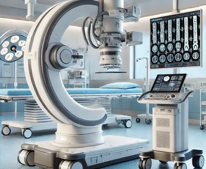
The arc in C is specialized medical equipment used in radiology and interventional procedures to obtain real-time X-ray images of the inside of the human body. It is a mobile device that enables radiological and fluoroscopic imaging. Its name derives from its C-shaped structure"which allows a wide range of movements and the acquisition of images from multiple angles and positions for capture specific anatomical views without moving the patient.
It is used to obtain X-ray and fluoroscopic images without having to move the patient to the radiology department. Therefore, diagnostics and procedures can be performed at the patient's hospital bedside or on the operating table during surgery. Its use is essential in areas such as surgery, orthopedics, traumatology, cardiology, neurology, urology and minimally invasive procedures.
Among the main advantages offered by the arc in Cis that facilitates diagnosisoffers a high precision and safety, y decreases the duration of surgical interventions in which the patient is under general anesthesia. In the following article, we analyze how a C-arm works, its parts, functions and main applications and uses. medical equipment.
How does a C-arc work?
The operation of a C-arc is the same as that of the X-ray machines conventional. Combine two main elements that work in an integrated manner How does this process work?
X-ray generator
The process begins with the X-ray tubelocated at one end of the "C" arm. This component emits a beam of radiation which passes through the patient's body. Collimators, which are adjustable devices on the tube, delimit the radiation field, ensuring that only the area of interest is irradiated. This not only improves image quality, but also minimizes radiation exposure to other areas.
When the X-ray beam passes through the patient's body, interacts with the different tissuesgenerating a phenomenon called differential absorption. The Denser tissues, such as bones, absorb more radiation. and are represented as white areas in the image. On the other hand, the soft tissues and air-filled areas allow the rays to pass through more easily, appearing in gray or black tones. This difference in absorption is what creates the contrast in radiological images.
Image detector or intensifier
At the opposite end of the X-ray tube is the image detector or intensifier. This component receives the rays that have passed through the patient and converts them into electrical signals. Modern detectors, called digital flat panel detectors, process these signals to generate high-resolution images. This advance has largely replaced traditional intensifiers, offering greater sharpness and less radiation exposure.
The signals captured by the detector are sent to a processing system that converts the data into digital images.. This software automatically optimizes parameters such as contrast, brightness and sharpness to ensure that images are clear and easy to interpret. These images are displayed in real time on monitors connected to the system, allowing the medical team to observe the area of interest while the procedure is being performed.
C-arc: Parts and functions
The C-arm in radiology consists of several parts that work together to provide high quality images in real time during medical procedures. Below are its main components and functions:
| Part | Description |
|---|---|
| C-shaped arm | Central structure connecting the X-ray tube to the detector. |
| X-ray tube | Located at one end of the C-arm, it emits the radiation beam. |
| Image detector | At the opposite end of the X-ray tube, it captures the radiation passing through the patient. |
| Mobile base | Wheeled structure that supports the equipment and facilitates its transport. |
| Control panel | Operational console from where the equipment parameters are adjusted. |
| Monitors | Screens connected to the image processing system. |
| Collimator system | Adjustable device located in the X-ray tube. |
| Cooling system | Components that dissipate the heat generated by the X-ray tube. |
1. "C" shape arm
It is the main structure that connects the essential components of the equipment, such as the X-ray tube and the imaging detector.
Functions:
- The C-shaped arm connects the X-ray tube, which is located at one end, to the image detector or intensifier, which is located at the opposite end, allowing a wide range of movement around the patient.
- Facilitates imaging from multiple angles no need to move the patient.
- Includes rotations in multiple planes: horizontal, orbital and verticalThis makes it possible to adapt to different types of procedures.
X-ray tube
This is the radiation generator located at one end of the C-arm.
Functions:
- Emits X-rays through the patient's body.
- Its intensity and duration are controlled to obtain quality images. while minimizing radiation exposure.
- Security is a key aspect in the use of the C-arm. These devices are designed to minimize radiation exposure for both the patient and the medical staff. They have specific systems that reduce scattered radiation and integrated dosimeters continuously monitor the delivered dose.
3. Image intensifier or digital flat detector
It is located on the opposite side of the X-ray tube, capturing the radiation passing through the patient.
Functions:
- Converts X-rays into visible images in real time.
- The state-of-the-art digital flat panel detectors offer higher resolution images and lower radiation exposure compared to traditional intensifiers.
4. Control Console
This is the external control panel operated by the radiological technician during diagnosis.
Functions:
- Allows adjustment of exposure parametersThe company has a wide range of products, such as time and intensity, among other aspects.
- Controls the movement of the arc and the orientation of the images.
- Saves and transmits the images obtained for further analysis. The data is stored in a PACS system (Picture Archiving and Communication System), allowing quick and easy access for further analysis.
3. Monitor
The C-arm includes one or more high-resolution monitors, usually in Full HD, which allow physicians to view images in real time during procedures. This screen is connected to the system, usually located near the surgical field.
Functions:
- Displays radiological and fluoroscopic images in real time so that physicians can be guided through the procedure.
- Some systems include dual monitors to compare images in real time with previous analyses.
6. Mobility system
It is a rolling base with lockable wheels or fixed support system on larger models.
Functions:
- Facilitates C-arm transport between different areas of the hospital.
- Allows you to position the equipment in a stable and safe manner around the patient.
7. Power generator
It provides the power needed to operate the X-ray tube and other system components.
Functions:
- Regulates the power supply to ensure consistent performance during use.
8. Image processing software
By means of a radiodiagnostic softwareThe computerized system manages the acquisition, processing and storage of medical images.
Functions:
- Improved image quality through techniques such as contrast adjustment and noise reduction.
- Allows measurements and annotations directly on the images.
9. Collimator system
It is the device located in the X-ray tube that is responsible for controlling the irradiated area to be analyzed or treated.
Functions:
- Adjusts the radiation field to focus only on the area of interest.
- Reduces unnecessary radiation exposure for both the patient and the medical staff.
10. Refrigeration system
The cooling system is the mechanism for dissipating the heat generated by the X-ray tube.
Functions:
- Maintains equipment temperature within safe operating limits.
- Prolongs X-ray tube lifetime.
Clinical uses and applications of a C-arm in radiology
The C-arm is a medical device widely used in radiology and interventional medicine due to its ability to generate real-time images with high precision. What are its main uses and clinical applications?
Orthopedic surgery
In the field of orthopedic surgery, the C-arm is essential for the precise placement of screws, intramedullary nails and plates used in orthopedic surgery. fracture treatment. It is also used for guiding fracture reduction or deformity correction procedures. Its ability to provide clear, real-time images allows the surgeon to visualize bone structures and ensure that implants are positioned correctly, reducing the risk of errors during surgery.
Spine surgery
In spinal interventions, the C-arm facilitates the precise placement of the fixation devices such as pedicle screws and spinal fusion brackets. In turn, it is also used in procedures such as the vertebroplasty. The real-time images it generates are crucial to avoid injury to sensitive nerve structures and to ensure a successful outcome.
Interventional radiology
The C-arm is an essential tool in interventional radiology, where it is used for guided procedures such as biopsies, drains and tumor ablations. It is also indispensable in angiographieswhere digital subtraction imaging (DSA) allows high-precision visualization of blood vessels. This equipment facilitates minimally invasive procedures, which require detailed, real-time imaging to ensure accurate results.
Interventional cardiology
In cardiology, the C-arc is used in procedures such as coronary angiographieswhich evaluates the circulation in the arteries of the heart. It is also key to the implantation of pacemakers and other cardiac devices. Thanks to the dynamic images it provides, physicians can perform complex procedures with greater safety and precision.
Vascular surgery
In vascular surgery, the C-arm allows detailed visualization of the vascular system, which facilitates procedures such as the stenting to repair aneurysms or the insertion of filters in the vena cava.
Urology
In urology, this equipment is used to guide procedures such as placement of ureteral catheters or nephrostomies. It is also useful in the percutaneous nephrolithotomywhere kidney stones are removed using minimally invasive techniques. Real-time imaging helps physicians locate specific structures and avoid damage to surrounding tissues.
Gastroenterology
In gastroenterologic procedures, the C-arm is used for inserting feeding tubes or drainsas well as for placing esophageal prostheses. This device is especially useful in delicate procedures where precision is crucial, such as in hard-to-reach areas within the gastrointestinal tract.
Neurosurgery
In neurosurgery, the C-arm is used for procedures such as the electrode placement for deep brain stimulation or minimally invasive spinal surgeries. The ability to generate highly accurate intraoperative images is critical for navigating complex structures of the nervous system and ensuring patient safety.
Oncology
In the treatment of cancer, the C-arm is a valuable tool for radiofrequency or microwave ablationswhere localized tumors are destroyed. It is also used for the placement of markers to guide radiation therapy. Its ability to generate precise images allows for accurate positioning of instruments in malignant tissues, optimizing treatment.
Traumatology
In emergency situations or in traumatology, the C-arc is used for evaluate complex fractures and guide reduction procedures. It allows to verify in real time the correct alignment of the bones, which is crucial to ensure the patient's functional recovery.
Emergency procedures
In emergency environments, this equipment is indispensable for the immediate evaluation of serious injuriesas major trauma, and for guiding critical procedures such as thoracic drainage. Its ability to generate immediate images allows physicians to make quick decisions and save lives in critical situations.
Dentistry and maxillofacial surgery
In dentistry and maxillofacial surgery, the C-arm is used for the dental implant placement and surgical planning in the mandibular region. Provides detailed images of the bony structures of the skull and jaw, ensuring accurate results.
Gynecology and obstetrics
In gynecology, this equipment is used for interventional procedures, such as the placement of intrauterine devices or catheters used in fertility treatments. Its use improves the accuracy of procedures in sensitive areas, increasing safety and effectiveness.
Conclusion
The C-arm stands out for its versatility, as it is used in multiple medical specialties. Its ability to provide real-time imaging facilitates decision-making during complex procedures, reducing errors and improving clinical outcomes. In addition, by enabling minimally invasive interventions, it contributes to faster patient recovery and greater efficiency in medical resources.


