por Luis Daniel Fernádez | Jan 16, 2025 | Equipment analysis
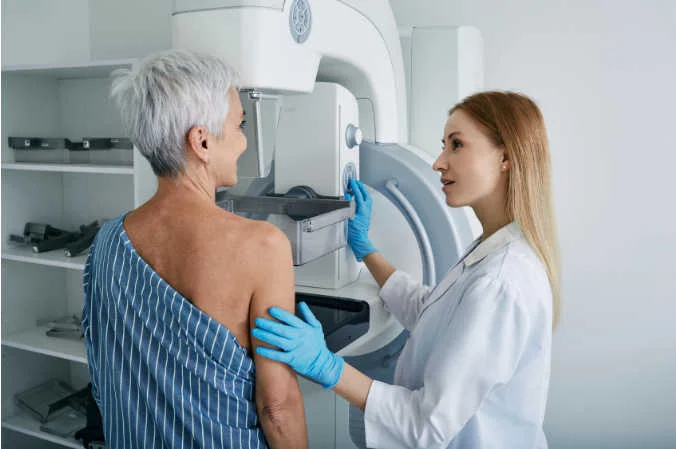
The mammography is a technique of diagnostic imaging which uses a system of low-dose X-rays to examine the inside of the breasts. This is a medical test that consists of performing a breast radiography. When performing a mammogram, a mammography machine is used. specific equipment: the mammograph.
It is a medical equipment that is specifically designed to capture X-ray images with a high resolution to detect signs and irregularities in breast tissue. The design and the different parts of a mammography equipment allow a minimum dose of radiation to be used during the test, making it an effective, fast and safe examination.
Health professionals use this test to look for early signs of disease in breast tissue. Among them, breast cancer. The mammography test is called mammogram and its main purpose is to detect abnormalities such as tumors, cysts or microcalcifications in the breast. We analyze, below, what mammography consists of, how the mammogram works and its different parts.
Mammography: What is mammography and types of mammograms?
The use of the mammograph is used as a screening tool for early detection of breast cancer in womenA mammogram is used both in women who have no symptoms and to diagnose the presence of abnormalities in women who notice breast irregularities. A mammography examination or mammogram exposes the woman to a small dose of ionizing radiation to generate medical images of the inside of the breasts. We can differentiate between two types of mammography:
Screening mammography
A screening mammogram is performed in women who have no signs or symptoms of breast cancer. This type of mammography should be performed periodically in women from the age of 40 as a form of prevention. By means of this diagnostic test, it is possible to detect irregularities in the breast tissue, such as tumors, cysts or microcalcifications. Screening for breast disease at early stages, especially breast cancer, provides a range of advantages:
- Allows the identification of tumors before they are palpable. or present visible symptoms.
- Enables treatment to be initiated in the early stagesbefore the disease has spread.
According to different studies, it has been proven that the screening mammography screening decreases breast cancer morbidity rates by detecting the disease at treatable stages, increasing the chances of successful treatment.
2. Diagnostic mammography
Diagnostic mammography is used when a woman presents symptomsas lumps, pain, discharge or changes in the skin of the breast. It is also used when an abnormality is detected on a screening mammogram or detection. This type of examination allows the affected area to be studied in greater detail and thus identify whether the breast condition is benign or malignant.
Mammograph operation
The medical equipment The mammogram is a specialized medical device that allows the analysis of breast tissue and the presence of abnormalities. This is specialized medical equipment that uses X-rays to generate medical images of the inside of the breasts. How a mammogram works consists of several stages:
Preparation of the patient
The process begins with the positioning of the patient in front of the mammograph. During the mammogram, a radiology professional will positions the breast on a flat platform of the mammography equipmentwhere the breast will be gradually compressed. The specialized technician will guide the patient to ensure proper posture and perform the medical test.
2. Breast compression
Once the breast is positioned, an adjustable compressor descends to press on the breast tissue gently, but firmly.
3. X-ray emission
The tube of X-rays of the mammogram emits a controlled beam of radiation passing through compressed breast tissue. This radiation is absorbed to a greater or lesser extent depending on the density of the tissue:
- The dense tissuessuch as tumors or microcalcifications, absorb more radiation. They appear clearer and brighter in the images.
- On the other hand, the fatty tissues absorb less radiation and appear darker.
4. Image capture
The radiation passing through the breast is captured by a detector which transforms the data into a digital image or radiographic film. Modern mammographs are often equipped with digital technology that allows images to be stored and processed on a computer.
Subsequently, these generated medical images can be integrated in the RIS system to automate the management of medical imaging data and information, facilitating its analysis and comparison with previous studies.
5. Variation of angles and views
To ensure a complete evaluation of the breast tissue, images are captured from different angles. The different perspectives help physicians identify abnormalities that may not be visible in a single view. The views that are analyzed in a mammography study are:
- Craniocaudal (CC)This is a top-down view.
- Mediolateral oblique (MLO)This type of slanted view allows a greater amount of breast tissue to be studied, especially that close to the axilla.
6. Image analysis
Once the images have been obtained, a specialized radiologist reviews the results for possible abnormalitiesas cysts, calcifications, tumors or suspicious tissue changes. Nowadays, digital images offer many advantages, since they allow adjusting contrast and brightness to improve image quality, obtaining a more efficient and accurate diagnosis.
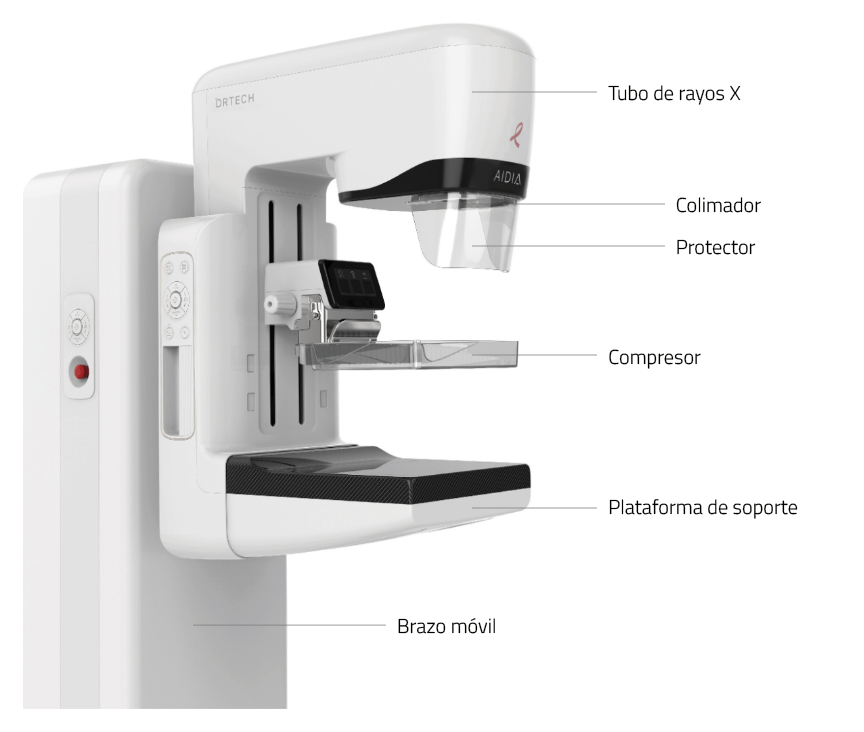
The mammograph: Parts and components
A mammogram is composed of several elements that work together to ensure clear and accurate images. Each component has a specific function that contributes to the quality of the diagnosis and the safety of the procedure. What are the main parts of a mammography machine?
X-ray tube
The X-ray tube is the component responsible for generating the X-ray beam that passes through the breast tissue. and subsequently produce high quality images. The mammograph uses a lower radiation doses than standard X-rays. This is because, since x-rays do not pass through this area easily, the mammography equipment is designed with two plates that compress and flatten the breast to separate the breast tissue. In this way, a higher quality medical image can be created and the amount of radiation during the exam can be reduced.
2. Compressor
The compressor is a movable plate that descends to press the breast against the mammography platform. Its function is to compress the breast tissue gently and firmly, providing the following advantages:
- Reducing the thickness of breast tissue to improve the visualization of internal structures.
- Minimizing X-ray scatteringThe image quality is improved.
- Avoid blurred images caused by the involuntary movement of the patient.
- Allowing the use of a lower dose of radiationmaking the procedure safer.
3. Support platform
The support platform is a flat surface on which the breast is positioned during mammography. It provides a stable and firm foothold, ensuring that the breast tissue is correctly positioned for sharp, detailed images.
4. Detector
The detector is the component that captures the radiation passing through the breast tissue and converts it into an image.. Depending on the type of mammograph, it can be of different types:
- DigitalX-ray: Converts X-rays into electronic data that is processed and stored in a computer, facilitating detailed and rapid analysis.
- Radiographic filmThis type of detector is used in analog mammographs, where the image is printed on a special film.
5. Collimator
The collimator is a structure that directs and confines the X-ray beam to the specific area of the breast that needs to be examined. This component prevents other areas of the body from receiving unnecessary radiation, making the procedure safer.
6. High voltage generator
The high-voltage generator is responsible for supplying the energy necessary for the X-ray tube to function correctly. It regulates the intensity and duration of the X-rays, adapting to the needs of each scan.
7. Control station
The control station is the panel or computer from which the technician operates the mammography machine. Allows you to adjust the parameters of the examinationIt also ensures that the procedure is performed in a precise and customized manner for each patient. It also ensures that the procedure is performed accurately and customized for each patient.
8. Positioning system
The positioning system includes mechanisms for adjusting the height, tilt and angle of the mammography machineThe system can be adapted to the physical characteristics of each patient. This system facilitates the imaging from different perspectivesobtaining a complete analysis of the breast tissue.
9. Image processing software
In digital mammographs, the digital mammogram processing software medical images is an advanced tool that improves the quality of captured images. Allows adjustment of contrast, brightness and other parameters to highlight specific details, as well as compare current images with previous studies, facilitating a more accurate diagnosis.
10. Security system
The mammogram is equipped with a safety system that ensures that radiation exposure is minimized and safe for the patient. In addition, some devices are equipped with sensors that automatically stop the process if a problem is detected technical or positioning.
Advantages of mammography
The mammograph is an essential medical device for the detection, diagnosis and follow-up of breast diseases, especially breast cancer. Its use not only allows early identification of abnormalities, but also contributes to more effective treatment planning. What are its main advantages?
Prevention and early detection of diseases
The mammograph is capable of identify abnormalities in breast tissue in early stages or even before symptoms and signs are visible. The early detection is key to significantly increasing the chances of successful treatment, as it allows the disease to be addressed before it develops to an advanced stage.
In turn, the periodic mammograms are performed is a fundamental strategy for the prevention of breast cancer in women. By detecting breast cancer in its early stages, it helps to reduce the mortality associated with this disease and improves the quality of life of patients.
Non-invasive, fast and safe procedure
Mammography is a non-invasive diagnostic procedure that uses a minimal dose of X-rays, meeting strict safety standards. The mammography exam is fast and efficient. It usually has a duration between 10 and 30 minutesdepending on the type of mammography performed:
- The screening mammogramsDuration: Its duration is between 10 and 20 minutes.
- The diagnostic mammogramsThey have a longer life, between 15 and 30 minutesThey include different views and images to analyze the area in a specific way.
High precision imaging
Modern mammographs, especially digital mammographs and those using 3D technology (tomosynthesis), provide high-resolution images that allow the breast tissue to be analyzed in great detail. This precision facilitates the detection of small or subtle irregularities and improves the differentiation between normal tissues and abnormalitiesreducing the probability of false positives or negatives.
Examination customization
The design of the mammograph allows tailoring the procedure to the individual characteristics of each patient. Exposure parameters, X-ray intensity, acquisition angle and compression level can all be adjusted. All this allows you to generate high quality medical images and optimize the patient experience.
Fast and efficient diagnostics
The mammogram streamlines the diagnostic process by generate medical images in a short period of time. In this way, when abnormalities are detected, physicians can immediately plan further studies and start treatment as soon as possible.
Multiple uses and clinical applications
In addition to being a key tool for the early detection of breast cancer, the mammogram has also other important applications:
- Monitoring of the evolution of oncological treatments.
- Performing image-guided biopsiesThis improves the accuracy of the procedure.
- Identification of benign changes or non-malignant disease in the breast tissue.
Conclusion
The mammograph is an advanced technological tool that combines precision, safety and efficiency for the detection and diagnosis of breast diseases.
If you need advice on radiodiagnostic medical equipment, at 4D we help you choose the most appropriate solution that suits your clinic's needs and budget. We have new and second hand equipment, as well as renting and leasing options. Contact us without obligation.
Contact 4D
Bibliography
American Cancer Society (n.d.).
Mammogram basics. Retrieved January 15, 2025, from
https://www.cancer.org/es/cancer/tipos/cancer-de-seno/pruebas-de-deteccion-y-deteccion-temprana-del-cancer-de-seno/mamogramas/conceptos-basicos-del-mamograma.html
RadiologyInfo.org (n.d.). Mammography. Retrieved January 15, 2025, from https://www.radiologyinfo.org/es/info/mammo
MedlinePlus (n.d.). Mammography. U.S. National Library of Medicine Retrieved January 15, 2025, from. https://medlineplus.gov/spanish/mammography.html
Centers for Disease Control and Prevention (CDC). (n.d.). Mammograms. Retrieved January 15, 2025, from https://www.cdc.gov/breast-cancer/es/about/mammograms.html
Revista Argentina de Mastología (2020). Importance of mammography in the early detection of breast cancer. Retrieved January 15, 2025, from https://www.revistasamas.org.ar/revistas/2020_v39_n141/06.pdf
Luis Daniel Fernandez Perez
Director of Diagximag. Distributor of medical imaging equipment and solutions.

por Luis Daniel Fernádez | Jan 7, 2025 | Equipment analysis
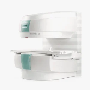
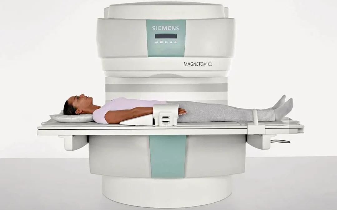
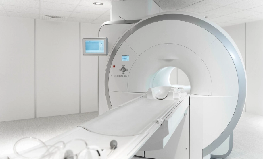
The magnetic resonance equipment MAGNETOM C!manufactured by Siemens brandoffers a compact and open design and incorporates advanced technology to achieve efficient image diagnosis. This medical equipment is a complete solution for healthcare facilities looking for an affordable, efficient and high-performance MRI equipment, suitable for a wide range of applications. wide range of medical applications. Its innovative design and advanced technology make it an essential tool for improving the quality of medical care and optimizing clinical resources. The following is an analysis of its technical characteristics, advantages and main clinical applications.
Technical characteristics of the Magnetom C!
The MAGNETOM C! 0.35T from Siemens is a team of magnetic resonance imaging designed to offer a balance between technological innovation, ease of use and patient comfort. Among its main features, we can highlight the following aspects:
Compact and open design
The design of the MAGNETOM C! is based on a C-shaped magnetThis design not only minimizes the size of the equipment, but also allows open access to the patient from 270°, which facilitates positioning and reduces the feeling of claustrophobia. This design not only minimizes the size of the equipment, but also allows open access to the patient from 270°, which facilitates positioning and reduces the feeling of claustrophobia. The 137 cm top opening and the vertical distance of 41 cm contribute to a more comfortable patient experienceespecially for those with anxiety or limited mobility.
In addition, its compact size makes it ideal for space-restricted facilitiesas it requires less than 30 m² for installation. This affordable and efficient design is especially suitable for small clinics and hospitals.
Magnetic field of 0.35 Tesla
The equipment uses a magnetic field of average intensity (0.35 Tesla)suitable for most diagnostic applications. This makes it possible to achieve a balance between image quality and operating costs, since no need for cryogenic cooling systems as the most powerful equipment.
The system includes an innovative hybrid shim mechanism, which combines active and passive methods to ensure homogeneity of the magnetic field. This ensures a homogeneous consistent image quality throughout the scan volumeeven in areas that are difficult to capture.
Multichannel technology
The MAGNETOM C! takes advantage of the multichannel technology to optimize imaging capabilities. Its ability to use up to four coils simultaneously allows detailed images to be captured and improves the efficiency of the scanning process. In addition, it is compatible with iPAT parallel acquisition technology, which speeds up scanning times without compromising image quality. This is particularly beneficial in long studies or with patients who have difficulty holding still during scanning.
Image quality and resolution
Although it is a mid-field device, the MAGNETOM C! offers a minimum resolution of 33 micrometers, which makes it possible to obtain sharp and detailed images that are suitable for a wide variety of diagnostics. It is also capable of performing 3D isotropic imagingthat can be reconstructed in any plane, facilitating the visualization and analysis of complex anatomical structures.
Technology and clinical application packages
The device comes equipped with a number of pre-installed applications, covering various medical specialties. Some of the highlights include:
- Neuro SuiteIt is designed for brain and spinal cord studies, with advanced sequences to detect tumors, lesions and neurological pathologies.
- Angio SuiteIt is used to perform angiographies without contrast, allowing to visualize arteries and veins in a precise and safe way.
- Cardiac SuiteCardiac morphology: Provides tools to evaluate cardiac morphology and function, as well as to diagnose congenital diseases.
- Body SuiteIt is a tool that optimizes the elaboration of abdominal and pelvic examinations, helping to identify pathologies such as tumors and liver diseases.
- Ortho SuiteIt allows the evaluation of joints and the spine, making it useful in musculoskeletal diagnostics.
- Pediatric SuiteProvides specific protocols for studies in children, adapting to their particular needs.
syngo MR software platform
The MAGNETOM C! uses the syngo MR software, a intuitive platform that facilitates equipment operation. This software automates complex tasksThe results are available immediately after scanning, including motion correction and image reconstruction. In addition, the Inline technology reduces the need for manual post-processing by performing automatic adjustments in real timeThe system can be used for pre- and post-contrast image subtraction. It also includes advanced 3D reconstruction toolsThe use of MPR (Multiplanar Reconstruction) and MIP (Maximum Intensity Projection), which are essential for analyzing anatomical and vascular structures.
Advantages offered by MAGNETOM C!
Siemens' MAGNETOM C! is an MRI system that provides a balance between advanced technology, accessibility and operational efficiency.
In the analysis of the equipment Magnetom C! doctor, these are its main advantages:
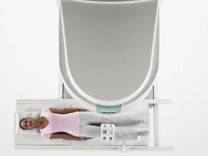
Open design: Accessible and comfortable for the patient
The "C" shaped design of the MAGNETOM C! provides a 270° open patient accessand therefore offers a less claustrophobic environment compared to traditional closed MRI systems. This significantly improves patient experienceespecially for those who suffer from anxiety or claustrophobia.
This MRI model has an adjustable tabletop and side access that can be used for facilitate precise patient positioning. This is very useful in interventional procedures or with patients who have limited mobility. Another advantage is that protocols are faster and minimize the time patients need to stay on the equipment. In addition, their capacity to support patients up to 200 kg makes it a suitable solution for a diverse population.
Source || Siemens healthineers
Wide range of clinical applications
The MAGNETOM C! is designed to cover a wide range of clinical applications, from neurology and cardiology to oncology, orthopedics and pediatrics. This makes it a comprehensive tool for medical centers serving different specialties. In addition, it offers customizable protocols that can be adjusted according to the specific needs of each case, guaranteeing precise diagnoses tailored to each patient.
Operational efficiency
The equipment is designed for optimizing clinical workflows. Its compatibility with parallel acquisition technology (iPAT) significantly reduces scanning times, allowing more patients to be seen in less time. Inline technology, which processes images in real time, eliminates the need for lengthy post-processing processes, delivering clinical results immediately after the study. In addition, its intuitive interface simplifies the use of the equipmentThis makes it possible to speed up the medical diagnosis.
Diagnostic quality at low cost
Although it is a mid-field device (0.35 Tesla), the MAGNETOM C! image quality excellent thanks to its advanced acquisition and processing technologies. This makes it an economical and effective option for most clinical needs.
Compared to high-field systems, this equipment has a more affordable initial costThe cryogen-free design reduces operating and maintenance costs, making it ideal for clinics and hospitals with tighter budgets. In addition, its cryogen-free design (no liquid helium) reduces operating and maintenance costs, making it ideal for clinics and hospitals with tighter budgets. more profitable in the long term.
Flexible installation
Thanks to its compact size, the MAGNETOM C! can be installed in confined spaces. In this way, only one space with dimensions of 30 m²This makes it ideal for clinics and hospitals with limited infrastructure. In addition, no need for significant modifications to the facilities and this facilitates its implementation even in small centers.
Profitability
The efficient design and versatility of the MAGNETOM C! offers an excellent return on investment. Its ability to address multiple medical specialties allows maximizing its use in a single facility, reducing the need to purchase additional equipment. The permanent magnet and simplified cooling technology guarantee a long equipment lifefor which the following are required minimum maintenance costs. Therefore, it is a solution that fits all types of medical centers.
Connectivity and data management
The equipment is compatible with the DICOM standardThis facilitates the transfer of images and data to other hospital systems for analysis and storage, such as the RIS system o PACS. With a storage capacity of up to 110,000 images, the MAGNETOM C! is a medical device that can store up to 110,000 images. enables management of large volumes of clinical data without interruptions.
Compatibility with interventional procedures
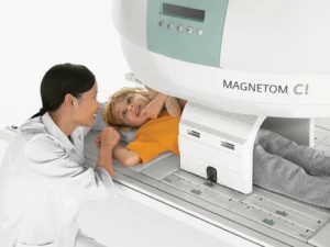
The open design of the MAGNETOM C!, coupled with the availability of wireless coils, makes it suitable for image-guided therapeutic procedures. During these procedures, the equipment provides real-time images, allowing physicians to make quick and accurate decisions.
Meet the 4D Médica Medical Team
Clinical uses and applications
The MAGNETOM C! is an MRI system designed to offer high quality diagnostic solutions in a wide range of medical specialties:
Source || Siemens healthineers
Neurological diagnosis
This medical equipment offers a detailed evaluation of the brain, spinal cord and nerve structures. This includes the detection of tumors, lesions, neurodegenerative diseases and congenital malformations. In addition, it enables specific studies, such as high-resolution imaging of the inner ear and cranial nerves, which are essential for complex diagnoses.
The system includes advanced protocols, such as 3D isotropic imaging, which allows detailed views in any plane. In turn, it is used to study specialized sequences for perfusion and diffusion studies, which are very useful in cases of stroke or ischemic pathologies.
Magnetic resonance angiography
The MAGNETOM C! allows the visualization of arteries and veins with advanced noncontrast angiographic techniquesThe system is an ideal option for patients with allergies or renal insufficiency. This equipment is capable of capturing detailed images of the vascular system, helping to diagnose conditions such as stenosis, aneurysms or thrombosis. To do so, it uses technologies such as time-of-flight (ToF) and phase-contrast to provide accurate and fast results in different anatomical areas.
Cardiological diagnosis
In the field of cardiology, the equipment facilitates the analysis of cardiac morphology, ventricular function and valves. It is particularly useful in the diagnosis of congenital diseases and cardiomyopathies. Thanks to its real-time dynamic imaging capability (TrueFISP cine), the MAGNETOM C! can capture the motion of the heart and provide critical information about its function.
Body images
The MAGNETOM C! stands out in the diagnosis of abdominal and pelvic diseases, including renal, hepatic and adrenal gland pathologies. Its high-resolution sequences, combined with advanced fat suppression techniques, allow clear visualization of internal organs. It thus aids in the identification of tumors, inflammation and other abnormalities.
Oncology
This equipment is an essential tool in oncology, since it provides detailed imaging for tumor detection and characterizationand for the follow-up of the response to treatment. Its ability to suppress fat signals and highlight soft tissues makes it ideal for visualizing lesions in different areas of the body. In addition, the dynamic protocols allow to evaluate the behavior of the lesions, which contributes to a more accurate diagnosis.
Orthopedic imaging
The MAGNETOM C! is widely used in the diagnosis of musculoskeletal and joint injuriessuch as tears, sprains and fractures. It is also effective in the evaluation of the spine and diseases such as avascular necrosis or bone tumors. Its high-resolution 3D sequences allow detailed views and multiplanar reconstructions, essential for a complete diagnosis.
Pediatric applications
The equipment offers specific protocols for pediatric studies, adapted to the needs of infants and neonates. This includes fast imaging for uncooperative patients and optimized sequences for developing tissues. It is useful for evaluating congenital malformations, tumors and epilepsy, as well as for cardiac studies in children.
Rehabilitation and sports imaging
In sports medicine and rehabilitation, this equipment is used for diagnose muscle, joint and tendon injuries. In addition, it allows dynamic analysis of moving joints, providing key information for treatment planning and evaluation of patient recovery.
Special applications
The open design and compatibility with specific accessories make the MAGNETOM C! an excellent choice for magnetic resonance-guided interventional procedures. In addition, its 270° access facilitates positioning of patients with special needsas those with claustrophobia or reduced mobility.
For more detailed information on the availability of our products, please contact us. magnetic resonance equipmentas well as leasing or financing options, you can contact 4D MédicaOur team will advise you and look for the best options for your clinic.
Conclusion
The MAGNETOM C! is an MRI model that combines a compact design with advanced technology to provide high-quality images in a variety of clinical applications. Its accessibility, ease of use and focus on patient comfort make it an essential tool for hospitals and clinics looking to enhance their diagnostic capabilities in an efficient and cost-effective manner.
If you want to learn more about magnetic resonance equipmentIf you have any questions, you can contact us and we will solve all your doubts so that you can choose the perfect medical equipment for your clinic or hospital.
Contact 4D
Luis Daniel Fernandez Perez
Director of Diagximag. Distributor of medical imaging equipment and solutions.
por Kiko Ramos | Dec 27, 2024 | Equipment analysis
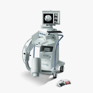
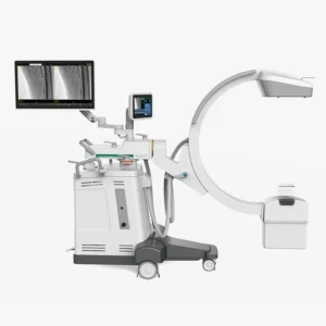
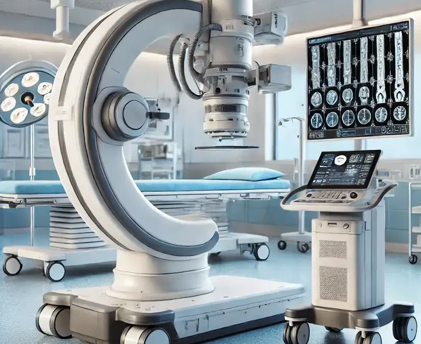
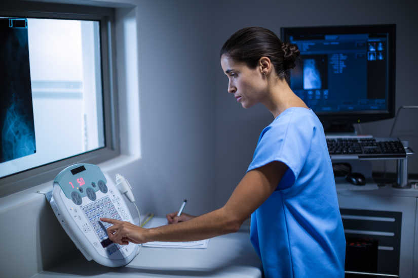
The arc in C is specialized medical equipment used in radiology and interventional procedures to obtain real-time X-ray images of the inside of the human body. It is a mobile device that enables radiological and fluoroscopic imaging. Its name derives from its C-shaped structure"which allows a wide range of movements and the acquisition of images from multiple angles and positions for capture specific anatomical views without moving the patient.
It is used to obtain X-ray and fluoroscopic images without having to move the patient to the radiology department. Therefore, diagnostics and procedures can be performed at the patient's hospital bedside or on the operating table during surgery. Its use is essential in areas such as surgery, orthopedics, traumatology, cardiology, neurology, urology and minimally invasive procedures.
Among the main advantages offered by the arc in Cis that facilitates diagnosisoffers a high precision and safety, y decreases the duration of surgical interventions in which the patient is under general anesthesia. In the following article, we analyze how a C-arm works, its parts, functions and main applications and uses. medical equipment.
How does a C-arc work?
The operation of a surgical C-arm is similar to that of the X-ray machines conventional. Combine two main elements that work in an integrated manner How does this process work?
X-ray generator
The process begins with the X-ray tubelocated at one end of the "C" arm. This component emits a beam of radiation which passes through the patient's body. Collimators, which are adjustable devices on the tube, delimit the radiation field, ensuring that only the area of interest is irradiated. This not only improves image quality, but also minimizes radiation exposure to other areas.
When the X-ray beam passes through the patient's body, interacts with the different tissuesgenerating a phenomenon called differential absorption. The Denser tissues, such as bones, absorb more radiation. and are represented as white areas in the image. On the other hand, the soft tissues and air-filled areas allow the rays to pass through more easily, appearing in gray or black tones. This difference in absorption is what creates the contrast in radiological images.
Image detector or intensifier
At the opposite end of the X-ray tube is the image detector or intensifier. This component receives the rays that have passed through the patient and converts them into electrical signals. Modern detectors, called digital flat panel detectors, process these signals to generate high-resolution images. This advance has largely replaced traditional intensifiers, offering greater sharpness and less radiation exposure.
The signals captured by the detector are sent to a processing system that converts the data into digital images.. This software automatically optimizes parameters such as contrast, brightness and sharpness to ensure that images are clear and easy to interpret. These images are displayed in real time on monitors connected to the system, allowing the medical team to observe the area of interest while the procedure is being performed.
Meet our 4D Medical equipment
C-arc: Parts and functions
The C-arm in radiology consists of several parts that work together to provide high quality images in real time during medical procedures. Below are its main components and functions:
| Part |
Description |
| C-shaped arm |
Central structure connecting the X-ray tube to the detector. |
| X-ray tube |
Located at one end of the C-arm, it emits the radiation beam. |
| Image detector |
At the opposite end of the X-ray tube, it captures the radiation passing through the patient. |
| Mobile base |
Wheeled structure that supports the equipment and facilitates its transport. |
| Control panel |
Operational console from where the equipment parameters are adjusted. |
| Monitors |
Screens connected to the image processing system. |
| Collimator system |
Adjustable device located in the X-ray tube. |
| Cooling system |
Components that dissipate the heat generated by the X-ray tube. |
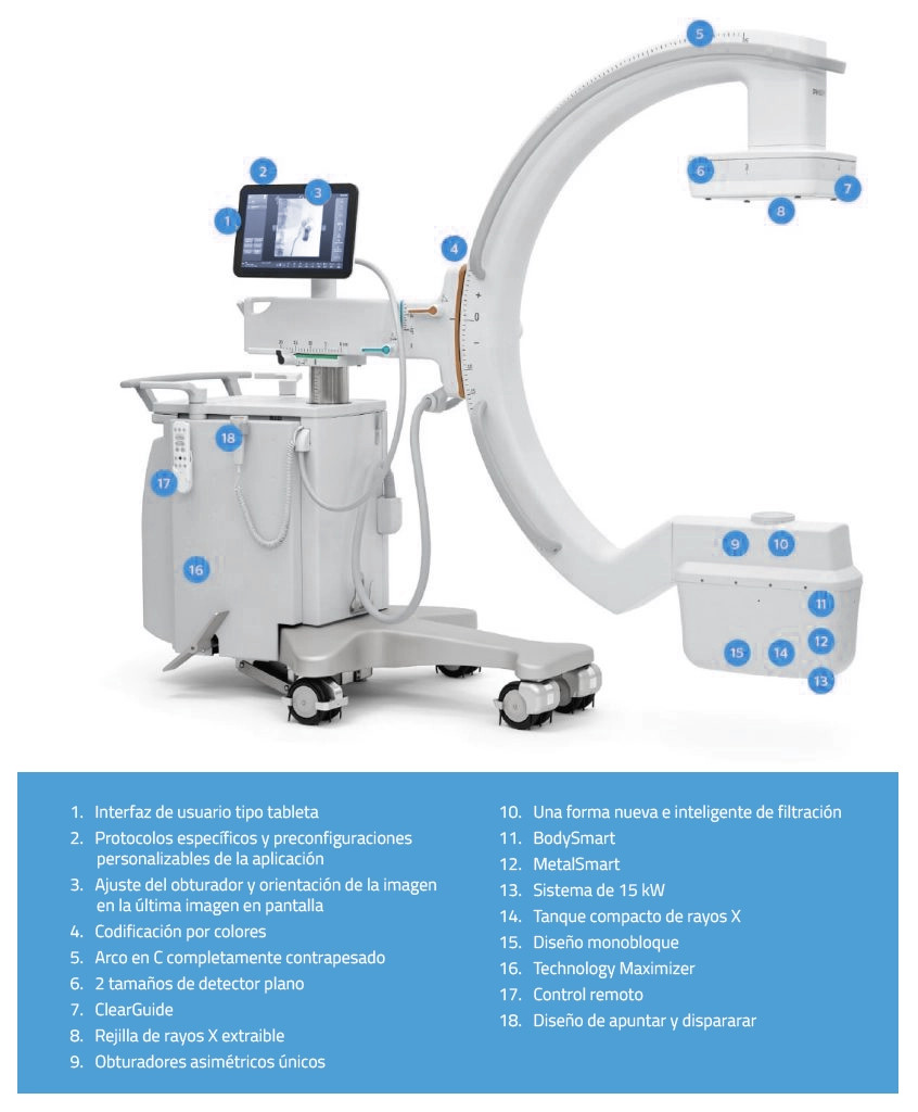
Parts of a C-arc
1. "C" shape arm
It is the main structure that connects the essential components of the equipment, such as the X-ray tube and the imaging detector.
Functions:
- The C-shaped arm connects the X-ray tube, which is located at one end, to the image detector or intensifier, which is located at the opposite end, allowing a wide range of movement around the patient.
- Facilitates imaging from multiple angles no need to move the patient.
- Includes rotations in multiple planes: horizontal, orbital and verticalThis makes it possible to adapt to different types of procedures.
X-ray tube
This is the radiation generator located at one end of the C-arm.
Functions:
- Emits X-rays through the patient's body.
- Its intensity and duration are controlled to obtain quality images. while minimizing radiation exposure.
- Security is a key aspect in the use of the C-arm. These devices are designed to minimize radiation exposure for both the patient and the medical staff. They have specific systems that reduce scattered radiation and integrated dosimeters continuously monitor the delivered dose.
3. Image intensifier or digital flat detector
It is located on the opposite side of the X-ray tube, capturing the radiation passing through the patient.
Functions:
- Converts X-rays into visible images in real time.
- The state-of-the-art digital flat panel detectors offer higher resolution images and lower radiation exposure compared to traditional intensifiers.
4. Control Console
This is the external control panel operated by the radiological technician during diagnosis.
Functions:
- Allows adjustment of exposure parametersThe company has a wide range of products, such as time and intensity, among other aspects.
- Controls the movement of the arc and the orientation of the images.
- Saves and transmits the images obtained for further analysis. The data is stored in a PACS system (Picture Archiving and Communication System), allowing quick and easy access for further analysis.
3. Monitor
The C-arm includes one or more high-resolution monitors, usually in Full HD, which allow physicians to view images in real time during procedures. This screen is connected to the system, usually located near the surgical field.
Functions:
- Displays radiological and fluoroscopic images in real time so that physicians can be guided through the procedure.
- Some systems include dual monitors to compare images in real time with previous analyses.
6. Mobility system
It is a rolling base with lockable wheels or fixed support system on larger models.
Functions:
- Facilitates C-arm transport between different areas of the hospital.
- Allows you to position the equipment in a stable and safe manner around the patient.
7. Power generator
It provides the power needed to operate the X-ray tube and other system components.
Functions:
- Regulates the power supply to ensure consistent performance during use.
8. Image processing software
By means of a radiodiagnostic softwareThe computerized system manages the acquisition, processing and storage of medical images.
Functions:
- Improved image quality through techniques such as contrast adjustment and noise reduction.
- Allows measurements and annotations directly on the images.
9. Collimator system
It is the device located in the X-ray tube that is responsible for controlling the irradiated area to be analyzed or treated.
Functions:
- Adjusts the radiation field to focus only on the area of interest.
- Reduces unnecessary radiation exposure for both the patient and the medical staff.
10. Refrigeration system
The cooling system is the mechanism for dissipating the heat generated by the X-ray tube.
Functions:
- Maintains equipment temperature within safe operating limits.
- Prolongs X-ray tube lifetime.
Clinical uses and applications of a C-arm in radiology
The C-arm is a medical device widely used in radiology and in the speciality of interventional radiology What are its main uses and clinical applications?
Orthopedic surgery
In the field of orthopedic surgery, the C-arm is essential for the precise placement of screws, intramedullary nails and plates used in orthopedic surgery. fracture treatment. It is also used for guiding fracture reduction or deformity correction procedures. Its ability to provide clear, real-time images allows the surgeon to visualize bone structures and ensure that implants are positioned correctly, reducing the risk of errors during surgery.
Spine surgery
In spinal interventions, the C-arm facilitates the precise placement of the fixation devices such as pedicle screws and spinal fusion brackets. In turn, it is also used in procedures such as the vertebroplasty. The real-time images it generates are crucial to avoid injury to sensitive nerve structures and to ensure a successful outcome.
Interventional radiology
The C-arm is an essential tool in interventional radiology, where it is used for guided procedures such as biopsies, drains and tumor ablations. It is also indispensable in angiographieswhere digital subtraction imaging (DSA) allows high-precision visualization of blood vessels. This equipment facilitates minimally invasive procedures, which require detailed, real-time imaging to ensure accurate results.
Interventional cardiology
In cardiology, the C-arc is used in procedures such as coronary angiographieswhich evaluates the circulation in the arteries of the heart. It is also key to the implantation of pacemakers and other cardiac devices. Thanks to the dynamic images it provides, physicians can perform complex procedures with greater safety and precision.
Vascular surgery
In vascular surgery, the C-arm allows detailed visualization of the vascular system, which facilitates procedures such as the stenting to repair aneurysms or the insertion of filters in the vena cava.
Urology
In urology, this equipment is used to guide procedures such as placement of ureteral catheters or nephrostomies. It is also useful in the percutaneous nephrolithotomywhere kidney stones are removed using minimally invasive techniques. Real-time imaging helps physicians locate specific structures and avoid damage to surrounding tissues.
Gastroenterology
In gastroenterologic procedures, the C-arm is used for inserting feeding tubes or drainsas well as for placing esophageal prostheses. This device is especially useful in delicate procedures where precision is crucial, such as in hard-to-reach areas within the gastrointestinal tract.
Neurosurgery
In neurosurgery, the C-arm is used for procedures such as the electrode placement for deep brain stimulation or minimally invasive spinal surgeries. The ability to generate highly accurate intraoperative images is critical for navigating complex structures of the nervous system and ensuring patient safety.
Oncology
In the treatment of cancer, the C-arm is a valuable tool for radiofrequency or microwave ablationswhere localized tumors are destroyed. It is also used for the placement of markers to guide radiation therapy. Its ability to generate precise images allows for accurate positioning of instruments in malignant tissues, optimizing treatment.
Traumatology
In emergency situations or in traumatology, the C-arc is used for evaluate complex fractures and guide reduction procedures. It allows to verify in real time the correct alignment of the bones, which is crucial to ensure the patient's functional recovery.
Emergency procedures
In emergency environments, this equipment is indispensable for the immediate evaluation of serious injuriesas major trauma, and for guiding critical procedures such as thoracic drainage. Its ability to generate immediate images allows physicians to make quick decisions and save lives in critical situations.
Dentistry and maxillofacial surgery
In dentistry and maxillofacial surgery, the C-arm is used for the dental implant placement and surgical planning in the mandibular region. Provides detailed images of the bony structures of the skull and jaw, ensuring accurate results.
Gynecology and obstetrics
In gynecology, this equipment is used for interventional procedures, such as the placement of intrauterine devices or catheters used in fertility treatments. Its use improves the accuracy of procedures in sensitive areas, increasing safety and effectiveness.
Conclusion
The C-arm stands out for its versatilityas it is used in multiple medical specialties. Its ability to provide real-time imaging facilitates decision-making during complex procedures, reducing errors and improving clinical outcomes. In addition, by enabling minimally invasive interventions, it contributes to faster patient recovery and greater efficiency in medical resources.
If you are a health professional and you are interested in to acquire a C-arc or any other radiodiagnostic equipment, our 4D team will contact you to advise you and find the best solution for your clinic or hospital.
Contact 4D
Kiko Ramos
CEO of 4D Médica. Expert in marketing and distribution of medical equipment.
por Luis Daniel Fernádez | Dec 18, 2024 | Equipment analysis
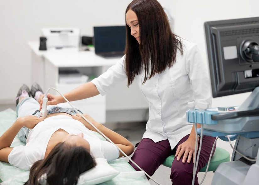
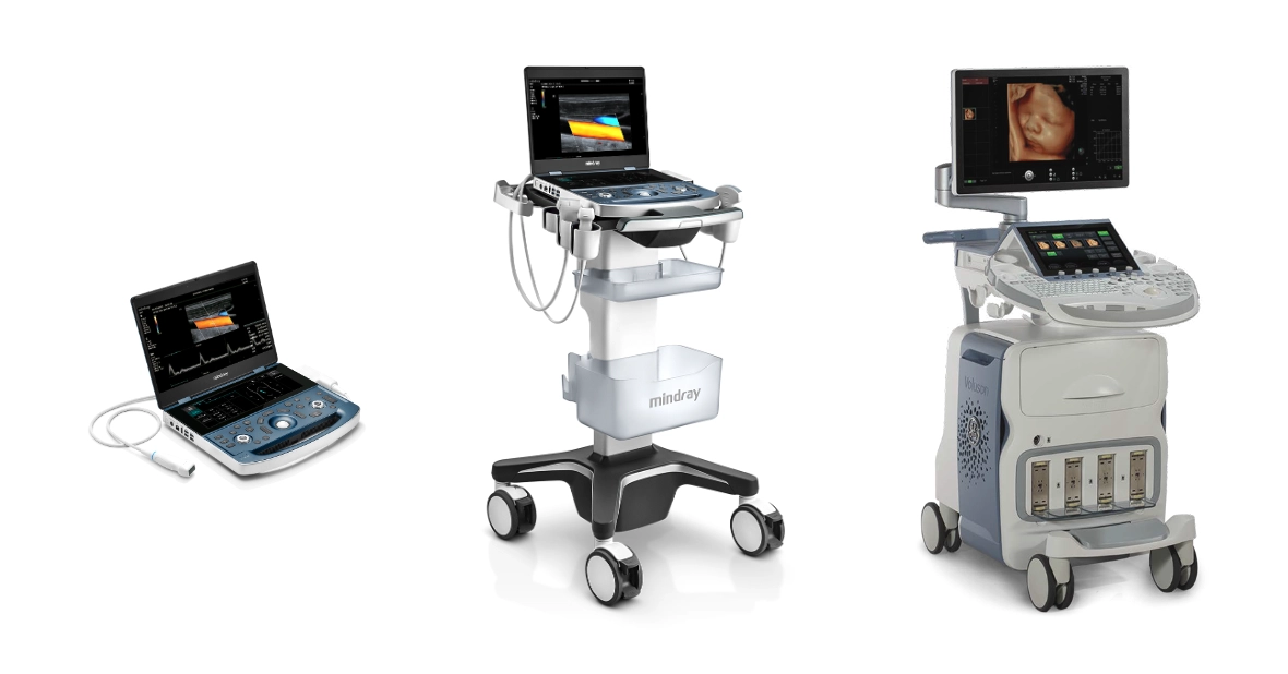
Ultrasoundultrasonography, also known as ultrasonography, is a non-invasive technique using ultrasound to obtain real-time images of the inside of the body. For this purpose, a medical equipment specific: the ultrasound scannerHow does it work and what types of ultrasound scanners are available on the market? We address this in the following article.
The ultrasound scanner: How does it work?
The ultrasound scanner is a medical equipment in the field of image diagnosis. It employs a device called a transducer which emits high-frequency sound waves, called ultrasound. These waves are inaudible to the human ear and travel through the different internal tissues of the body. At the moment when the waves encounter the various organs and structures, it is when are reflected as echoes. These echoes are picked up by the transducer and generate the medical images that can be displayed on a screen. These images are known as ultrasound scans and allow professionals to evaluate different tissues and internal organs of the organism.
In the realization of a ultrasoundis used, a transducer that glides over the skin in the area to be analyzed. This device is coated with a conductive gel that facilitates the transmission of ultrasound waves. It has the function of eliminating the air that exists between the skin and the transducer, helping to improve the quality of the images. In an ultrasound scan, the following can be obtained still images and also allows to observe the movement in real time. It is an essential medical equipment in medicine that has the function of analyzing the state of organs such as the heart or blood flow.
Meet our 4D Medical equipment
Parts of an ultrasound scanner
An ultrasound scanner consists of the following components:
| Parts of an ultrasound scanner |
Description |
| Transducer or probe |
Device in charge of emitting and receiving ultrasonic waves. |
| Monitor |
Screen where the images generated by the ultrasound scanner are displayed. |
| Control panel |
Interface with buttons and controls to adjust parameters and settings. |
| Central processing unit |
Processor that handles the data and generates the ultrasonic images. |
| Storage system |
Allows to save images and data obtained during diagnosis. |
| Power supply |
Provides electrical power to the device. |
| Software |
Program that controls the operation of the ultrasound scanner and processes the images. |
| Handles and wheels |
Facilitate the mobility of the equipment within the hospital or clinic. |
| Ports and connections |
They allow the connection of accessories and additional devices. |
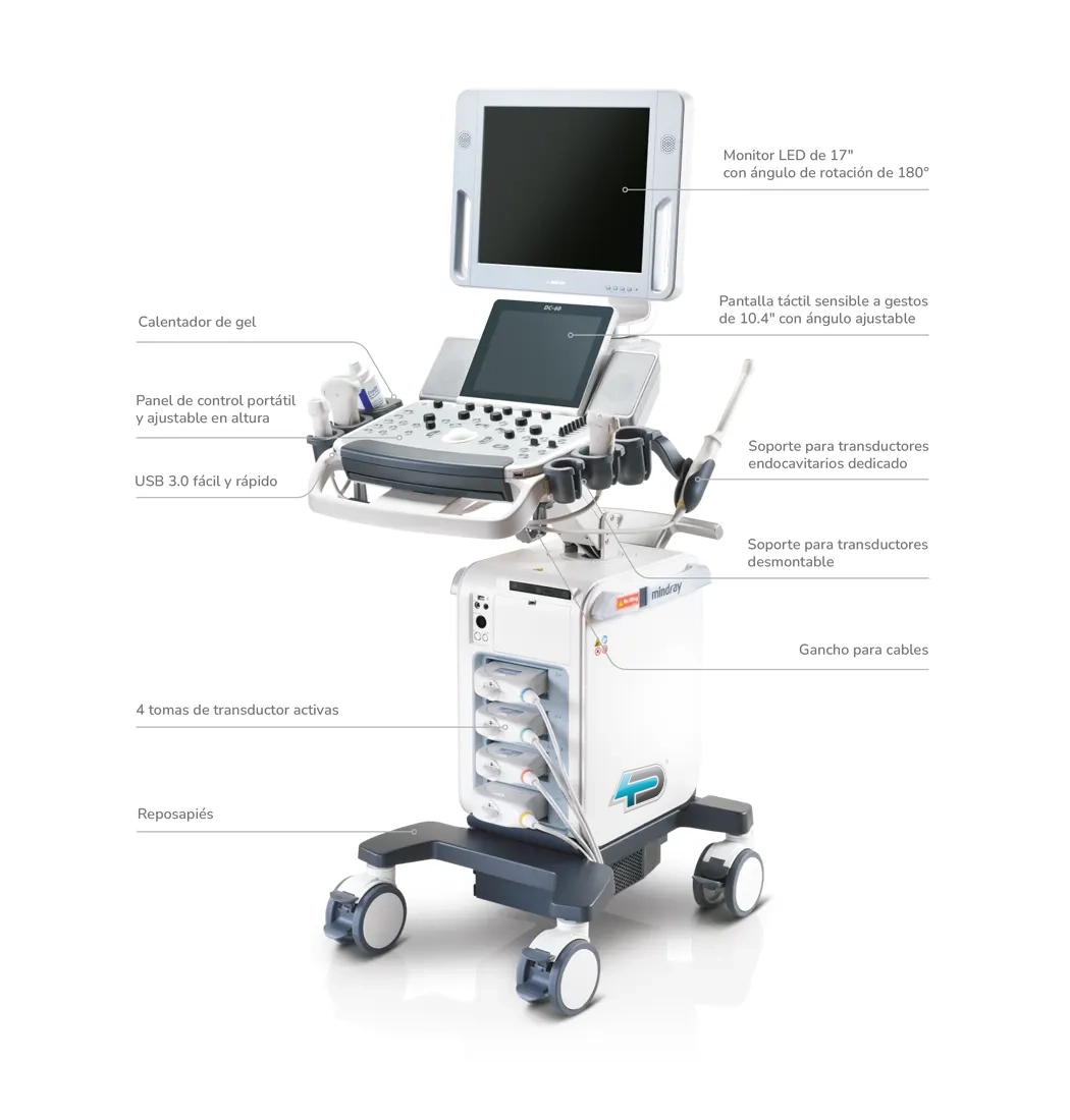
Detailed image of the parts of an ultrasound scanner
Transducer or probe
It is the main part of the device, responsible for transforming electrical signals into ultrasound waves. They are made of piezoelectric material and function as ultrasound emitters and receivers. There are different types of transducers:
Depending on its use
- LinearThey are used for superficial and vascular studies. They generate rectangular images and use high frequencies, since they do not require much penetration, being useful in the exploration of ligaments, tendons, muscles, thyroid, scrotum, breast and superficial vessels.
- Curved or convexThey have a curved shape and produce trapezoidal images. They are used with low frequencies because they are designed to explore deep structures, as in obstetrics and abdominal studies in general.
- Endocavitary or intracavitaryThey can be linear or convex. Their frequency varies according to the required penetration. They are used in intravaginal and intrarectal studies, for gynecological or prostate examinations.
- SectorialThey are a variant of the convex transducers and offer triangular or fan-shaped images. They use frequencies similar to those of curved transducers and allow an intercostal approach, so they are used in cardiac and abdominal studies.
According to frequency
- High frequency (up to 15 MHz)They are used to explore small and superficial structures.
- Low frequency (approximately 2.5 MHz)They are used for ultrasound scans that require a greater depth of penetration.
Monitor
Displays the images generated by the processing unit.The image is displayed on the monitor, so that professionals can observe and evaluate the state of the different anatomical structures in real time. Most current monitors can reproduce images in grayscale and color.
Control panel
It is located in the front part of the ultrasound scanner and allows the ultrasound specialist to make various adjustments to the equipment configuration. It allows to modify the brightness, the sharpness of the images and the frequency of the sound waves. In addition, it also allows to configure the necessary parameters to carry out the type of ultrasound that the patient requires.
Central processing unit
It is the component that receives the information provided by the probe. It converts the signals into electrical impulses and generates the image of the anatomical part of the area to be analyzed.
Storage system
It is the internal element that allows to save images and patient's data for further analysis. It can consist of an internal memory, USB or be connected to a PACS system (Picture Archiving and Communication System).
Power supply
Provides power to the ultrasound machineThe power supply is provided either by alternating current or by rechargeable batteries in the portable models.
Software
It is essential for processing ultrasound signals and generating medical images. It can include specific modules for different types of studies, such as cardiology or gynecology, among other areas.
Handles and wheels
These elements facilitate handling and transport of the equipmentespecially in the case of mobile ultrasound scanners.
Ports and connections
This type of components included in the ultrasound scanners are used for connect multiple probes, USB devices or DICOM interfaces to share images.
Types of ultrasound scanners
Having analyzed the operation of an ultrasound scanner and its main components, we can differentiate between different types of ultrasound scanners:

Imaging technology
1. 2D ultrasound scanners
- These are the most common and basic models. Generan two-dimensional images in real timeThey are widely used in the obstetrics area, to perform general and abdominal studies.
- Main applicationsBasic analysis, pregnancy control and organ evaluation.
2. 3D ultrasound scanners
- Allow display three-dimensional structures in real timeproviding greater detail. They are useful for creating more accurate images of fetuses and studying structural abnormalities.
- Main applicationsThey are used in advanced obstetrics and for surface studies of organs and tumors.
3. 4D ultrasound scanners
- They add the time dimension to 3D imagesallowing to see the movement in real time. It is especially useful in the obstetrics area to see fetal movements.
- Main applicationsObstetrical diagnosis and dynamic studies of joints.
4. Doppler ultrasound scanners
- They use the Doppler effect for assessing blood flow in vessels and organs. There are different models and variants:
- Color DopplerThey offer a color representation of the blood flow.
- Pulsed Doppler technologyThey provide a more detailed analysis of blood flow velocities.
- Continuous DopplerThey measure very fast flows.
- Main applicationsThey are used for vascular, cardiac and circulatory studies.
5. Tissue Doppler Ultrasound Scanners
- They are in charge of making a specific evaluation of the movements of the heart tissues and blood flow.
Mobility
1. Portable ultrasound scanners
- They are small and lightweight devicesThey are ideal for home transport, emergency or remote areas. There are multiple versions that include advanced technologies, such as 2D ultrasound, Doppler, etc.
- Main applicationsThey are used for emergencies and ICU, mobile clinics and medical visits to remote areas.
2. Trolley or console ultrasound scanners
- They are larger and more robust models. They have a fixed console that offers a variety of functions and high-resolution imaging options.
- Main applicationsThey are used in hospitals and specialized clinics.
3. Wireless ultrasound scanners
- They are connected to mobile devicesThe medical imaging systems, such as tablets or smartphones, through applications. They are characterized by high portability and immediate access to the generated medical images.
- Main applicationsThey are used in sports medicine, emergencies and telemedicine.
Clinical Specialty
1. Obstetrics and gynecology
- This type of transvaginal ultrasound scanners are specialized in the visualization of the fetus, uterus and ovaries of women.
2. Cardiac (Echocardiograms)
- They are designed to evaluate the structure and heart function, valves and blood flow.
Vascular
- They are used for analize arteries and veinsmeasuring the flow and detecting obstructions or thrombi.
4. Musculoskeletal and Physical Therapy
- Allow visualizing muscles, ligaments, tendons and joints. These physiotherapy ultrasound scanners are used in sports medicine to detect injuries or to analyze the recovery from an injury.
5. Abdominals
- They are oriented to the study of abdominal organs like the liver, kidneys, spleen or pancreas.
6. Neurological
- They are used for assessing the brainespecially in neonates.
7. Urological
- These devices are designed to examine the kidneys, bladder and prostate of the male.
8. Endoscopic
- They combine ultrasound with endoscopes to obtain internal images of the digestive tract or areas of difficult access.
Resolution and advanced technology
1. High resolution
- This type of medical equipment offers images of the highest qualityIt is therefore especially useful in complex applications.
2. Ultrasound scanners with Artificial Intelligence (AI)
Type of purchase
1. New ultrasound scanners
New ultrasound scanners are newly manufactured, previously unused ultrasound machines with the latest technology upgrades and full manufacturer's warranties. They feature the following characteristics:
- State-of-the-art technologyThey incorporate the latest innovations in imaging, such as advanced Doppler, elastography, 3D and 4D ultrasound and even artificial intelligence.
- Full warrantyThey offer extensive warranties that are backed by the manufacturer, generally from 1 to 5 years.
- CustomizationYou have the possibility to configure the equipment according to your specific needs, including transducers and software.
- Longer service lifeSince they have no previous use, their potential useful life is longer, especially if proper maintenance is carried out.
- Certifications and technical supportThey comply with all current quality and medical safety standards. In addition, they have specialized technical support.
2. Second-hand or opportunity ultrasound scanners
The used ultrasound scanners are previously used ultrasound devices that have been reconditioned or overhauled to ensure their functionality before being sold again. These devices may come from clinics, hospitals or doctors' offices that have refurbished them for newer models or no longer need them. Compared to new models, they have the following features characteristics:
- Technical reviewBefore being sold, ultrasound scanners undergo a series of quality tests to ensure that they are functioning properly. These may include repairs, cleaning, calibration and software upgrades.
- Reduced priceThey are less expensive than new equipment, which makes them attractive for small clinics, independent physicians or institutions with limited budgets.
- Variety of modelsYou can find from basic ultrasound scanners to advanced equipment with technologies such as Doppler or 3D.
- Limited WarrantySome suppliers offer warranties, but these are usually shorter than those for new equipment.
- Variable statusThe performance and service life of used ultrasound scanners will depend on how well the device has been maintained during previous use.
Conclusion
The ultrasound scanner is a medical equipment that is widely used in the field of diagnostic imaging to perform one of the most popular medical tests: ultrasound. Depending on the technology, mobility, medical specialty and type of purchase, different types of ultrasound scanners can be found.
There is a wide range of ultrasound scanners on the market that adapt to each of the medical needs. If you need more information, contact us and from 4D Médica we will offer you personalized advice so that you can choose the most suitable ultrasound scanner for your center.
Contact 4D
Luis Daniel Fernandez Perez
Director of Diagximag. Distributor of medical imaging equipment and solutions.
por Luis Daniel Fernádez | Dec 13, 2024 | Equipment analysis
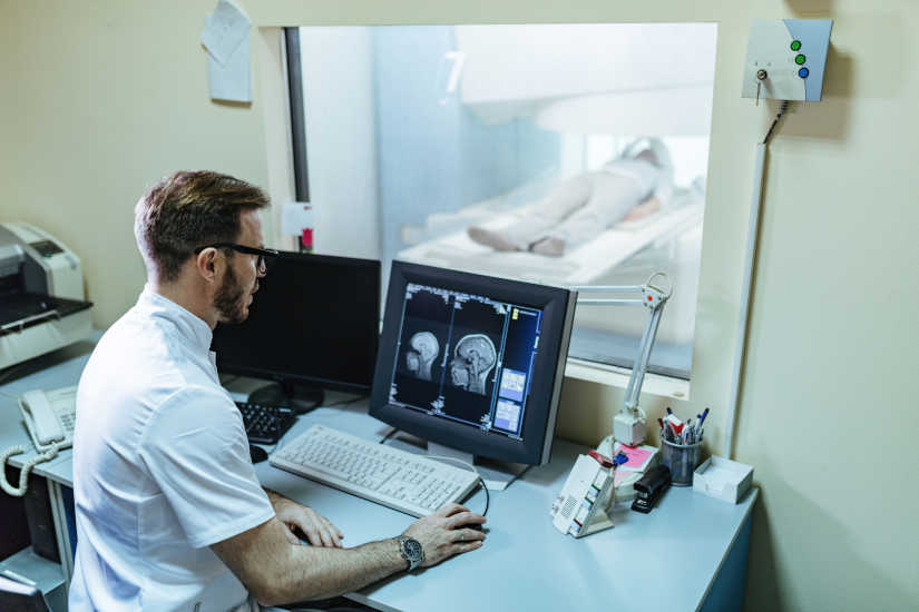
The technology has had a significant impact on the healthcare system, especially in the radiology area. In recent years, one of the most relevant changes following the advent of the Internet has been the use of computerized systems in the field of image diagnosis. This has allowed the development of a digital imaging department where medical information can be managed and stored conveniently and securely.
In a digital imaging department, we can distinguish three fundamental tools: the PACS system, the RIS system and the HIS system. In the following article, we analyze what the PACS system is, how it works and its relationship with the RIS and HIS system.
What is the PACS system in radiology?
The term PACS stands for Picture Archiving and Communication System, which refers to Image Archiving and Communication System. This is a computer software used in the radiology area for the following purposes store, manage, present and share medical images and diagnostic procedure reports electronically.
Before the advent of the PACS system in radiology, the images generated after diagnostic examinations were stored in a physical format, mainly as radiographic films. Therefore, from the time the medical test was performed, there was a long process until the final image was obtained. With digitization, it is now possible to resort to a AI software for the different medical teams to obtain an accurate faster and more efficient access to informationwhich will allow optimize workflow in clinical practice.
How does the PACS system work?
A PACS system consists of a series of mechanical and electronic components which are connected to each other by a copper or fiber optic communication network. Specifically, we can differentiate between four main components:
- Image acquisition hardware
- Workstations for image interpretation and review
- Servers for storage and transmission of images
- Network for data transmission
All these elements work in an integrated manner to allow medical images to be captured, stored, distributed and displayed digitally. Through the use of this network, the graphic information generated in different studies, such as a CT scan, is transmitted to the magnetic resonance imaging o TAC.
How does this process unfold?
First of all, data from the system servers is passed to the archiving drives. Subsequently, they are distributed to the stations where radiological physicians review the generated medical images and also to the teleradiology serverswhich allow access to the archive through the Internet.
With a digital radiology PACS system, you can view images remotely from any medical department, office or externally. To do so, health care personnel have special identification codes which allows them to access diagnostic tests for each patient.
The DICOM medical imaging communication standard
For information and images to flow through the PACS system components, it is necessary to comply with the DICOM medical image communication standard. DICOM stands for Digital Imaging and Communications in Medicine and is a standard for the communication of medical images. standard for digital storage and transmission of medical images and related patient information.
It is responsible for define the file format and structure and, in turn, includes a communications protocol to facilitate connectivity between medical devices and systems. However, it should be noted that the majority of modern devices and medical equipment current DICOM images are produced.
Advantages of using a PACS system in radiology
We analyze the main advantages offered by a PACS system in the management of radiological images:
Improved workflow in radiology departments
Radiologists and medical teams involved in the diagnostic imaging process can access and review digital images from any workstation on the hospital's network or remotely through the web server. This allows rapid consultation of studies and collaboration between physicians and specialists.
Error reduction
As the format of medical images is no longer physical, eliminating the possibility of duplicate diagnoses and also reduces both the risk of loss as the damage of the generated medical images.
Integration with other IT systems
One of the main advantages of the PACS system is that it allows the integration with other IT systems that can be used in health careThe RIS (Radiological Information System) and HIS (Hospital Management Software).
Capacity to store large volumes of data
Not only is it essential for clinical management and patient care, being able to store large volumes of medical imaging data is a key aspect for research and education in the area of health and medicine. In this way, researchers can access image databases for studies and students in training can use many of the images as educational material.
More accurate and detailed diagnosis
The use of the PACS system provides a more detailed reading of the diagnoses. This is mainly because the images are reviewed on high-resolution monitors and can be manipulated more accurately, which helps to detect abnormalities present in the image more quickly and accurately.
Saving time and resources
Another of its advantages is that it offers a time saving and a decrease in the workload of the staff.The cost of printing X-rays and other radiological elements was also reduced. At the same time, waiting times and resources at the hospital level are reduced.
Relationship between the PACS, RIS and HIS system
PACS, RIS and HIS are three systems key components in the digital health informatics ecosystem. Their interrelation is essential for the efficient functioning of the healthcare services of any clinic, health center or hospital. While the PACS system in radiology is used to manage, store and share images of the different diagnostic imaging procedures, the RIS and HIS system have other functions. What is each used for and what is the relationship between them?
The RIS system
The RIS system or Radiology Information System, is the program that runs the digital radiology department. It is a software that contains all the information of the radiology area and hospitals, thus enabling manage information and processes related to diagnostic imaging services.
Functions performed
- Scheduling of appointments and studies
- Order generation
- Recording of results with the generated medical images
- Workflow management in the radiology department
The HIS system
As for the Hospital Information System (HIS), it is a system of hospital information system. By using it, all the data are stored in the data related to the management and administration of a hospital. It is designed to manage all areas involved in the operation of a hospital from a single platform.
Functions performed
- Management and scheduling of medical appointments
- Patient care: Administration of patients' medical records and results of medical examinations performed.
- Human Resources
- Billing
- Monitoring the quality of medical care
Interaction of PACS, RIS and HIS systems
- HISThe central system that coordinates and stores all patient information in a clinic or hospital facility, including demographic, clinical and financial data.
- RISIt communicates with the HIS system to obtain relevant patient information and to manage the radiology area. It is used to schedule radiological procedures requested from other areas of the hospital.
- PACSRIS-PAC: Works hand in hand with RIS to store and manage the medical images generated by the requested studies. The RIS-PAC interaction allows the report to be presented in both systems so that each report appears linked to the images of the study performed.
Conclusion
The PACS system is a fundamental tool in the radiology area to be able to store and manage medical images digitally. All of this helps to improve healthcare and drive faster, more detailed and accurate clinical diagnosis.
If you need more information about our imaging solutions, just contact us and our staff will give you personalized advice.
Contact
BIbliography
Clínica Universidad de Navarra (n. d.). PACS. Medical dictionary. Retrieved from
https://www.cun.es/diccionario-medico/terminos/pacs
Ochoa, P. J., Murillo, M. R., & Torres, J. A. (2004). PACS system (picture archiving and transmission system). Anales de Radiología de México, 3(3), 153-162. https://www.analesderadiologiamexico.com/previos/ARM%202004%20Vol.%203/ARM_04_3_3_Julio-Septiembre/arm_04_3_3_153-162.pdf
López-Arroyo, A., Villarreal-García, A. J., & López-Arroyo, S. (2005). The DICOM format and PACS systems in medical imaging. Gaceta Médica de México, 141(5), 477-485. Retrieved from https://www.scielo.org.mx/pdf/gmm/v141n5/v141n5a11.pdf
Clinic Cloud (n. d.). DICOM format: what it is and how this standard works in medical imaging. Retrieved from https://clinic-cloud.com/blog/formato-dicom-que-es-estandar-imagenes-medicas
Luis Daniel Fernandez Perez
Director of Diagximag. Distributor of medical imaging equipment and solutions.
por Luis Daniel Fernádez | Nov 25, 2024 | Equipment analysis
Technology is becoming increasingly important when it comes to storing and managing different data and resources. In the field of medicine, we can highlight the RIS management system for diagnostic imaging. This is a type of specialized software used in the radiology area and in other medical fields to manage information and processes related to the services provided by the image diagnosis. In the following article, we analyze how it works, its main features and advantages.
What is the RIS management system for diagnostic imaging?
The RIS management system is responsible for automating the management of medical imaging data and information. It works like a hospital information system (HIS), but the main difference is that it is specifically tailored to radiology departments in clinics, hospitals and healthcare centers.
It is called RIS (Radiology Information System) and represents a key part of the IT infrastructure in radiology departments, clinics and hospitals. A radiodiagnostic software is a tool that includes a multitude of functions in a single centralized platformfrom manage patient data and history, store medical images and create customized reports. Therefore, it stands out as a solution that helps to improve workflows and optimize medical imaging processes.
Main features and functions of the RIS system
How does the RIS system work? We analyze the main features and functionalities it offers:
Patient registration
Firstly, the RIS system is used to register the patients to be attended. For this purpose, the different data to create your medical record: the personal information of contact, the medical history and the insurance information.
Appointment scheduling
Once the patients are registered in the system, they can be scheduling appointments for diagnostic imaging tests. From radiographs, computed tomography or CAT scans, magnetic resonancesetc. The software organizes and prioritizes orders according to urgency, equipment and personnel availabilityoptimizing the management of time and available resources.
Storage and tracking of medical images
Radiologists can attach the results of the images generated after the medical tests directly to the patient's fileThis speeds up the availability of the studies. At the same time, it also allows include data related to medical examinationssuch as reports and diagnostic information.
Patient follow-up and test management
The RIS system makes it possible to perform the follow-up of the patient's treatment and of the examinations carried out through the system. In this way, the complete medical history can be accessed and patient information can be checked for necessary updates during the diagnostic process.
Workflow tracking
Allows you to track each stage of the process, from the initial request to the generation of the final reportThe system ensures efficient and uninterrupted execution. Another aspect to highlight is that improves collaboration between different medical teams who work in patient treatment, such as radiologists, technicians and medical specialists.
Report generation
Radiologists can writing and sharing diagnostic reports based on processed images. The reports are securely stored and made available to physicians and also to authorized patients. The results are generated digitally, but can also be sent by e-mail and fax, as well as exported for printing on paper. Using the RIS system, different statistical reports can be produced, either for specific examinations, individual patients or groups of patients.
Data analysis and statistics
The system produces reports and statistics on workflows, volumes of studies performed and equipment performanceThe results of this study will facilitate administrative decision making and increase the efficiency of diagnostic imaging services.
Data storage and security
All information, including images, reports, and financial records, is stored in secure databases. This helps to ensure the compliance with medical and privacy regulationssuch as GDPR in Europe or HIPAA in the United States.
Billing and administration
Another of its functions is that automates the creation of invoices related to exams performed. By integrating payment and insurance records, financial management processes can be simplified.
What are the advantages of RIS for diagnostic imaging?
The RIS management system offers numerous advantages, mainly in terms of efficiency, accuracy and quality of service in the field of radiology. We explain its main benefits in the medical field:
Workflow optimization
Allows you to manage all stages of medical diagnosisfrom the request to the delivery of reports. This helps to improve organization and reduce delays that may arise. At the same time, automated appointment scheduling ensures that the efficient use of time and resources.
2. Accuracy and security of data
Reduces the occurrence of errors by centralizing patient information, as test results are located on a single platform. On the other hand, by complying with data security regulations such as HIPAA and GDPR, the medical information included in the RIS system is kept confidential.The patient's data is processed correctly.
3. Quick access to information
Physicians, radiologists and technicians have immediate access to patient records and studiesThis streamlines clinical decision making. And not only that, the system usually includes a integration with cloud-based solutions. In this way, the medical team can remotely access information from anywhere, anytime.
4. Integration with other medical systems
It works in conjunction with other medical systems: both PACS and HIS. On the one hand, the PACS system is used to manage the long-term storage of both images and patient information, and HIS systems are hospital information software used in the management of clinics and hospitals. Therefore, the integration of these systems into the RIS system makes it possible to create a complete healthcare ecosystem.
5. Improved patient care
Offers a agile, comprehensive and seamless patient care experience. Among its advantages is the reduction of waiting times in treatment planning and diagnosis, the results are available more quickly and reduce the administrative burden to be carried out by professionals and patients.
6. Cost reduction
In addition to optimizing the work process, helps reduce costs and increase profitability. It eliminates the need to create paper documentation and reduces administrative errors, thus optimizing billing processes and scheduling of medical services.
Conclusion
The RIS management system is an essential tool for optimizing administrative and clinical processes in radiology and other areas of diagnostic imaging. The use of radiodiagnostic software helps to increase efficiency, service quality and patient care.
Luis Daniel Fernandez Perez
Director of Diagximag. Distributor of medical imaging equipment and solutions.