Computed Radiography: How it Works and Workflow
Computed radiographyalso known as CR (Computed Radiography), is a technique of image diagnosis which represents a transition between conventional radiology and current digital technologies. Instead of using traditional radiographic film, CR uses photoluminescent phosphor plates that store the energy of the X-rays. This energy is then released and converted into a digital image by a laser readout process.
This system makes it possible to digitize radiographic images without the need to completely transform the X-ray service infrastructure. It is therefore considered to be a intermediate solution between analog and direct digital technology (DR). It is especially useful in clinics or centers seeking to modernize their equipment without making investments as high as those required by DR. In turn, computed radiography can also be used to facilitates the storage, archiving, distribution and analysis of images in digital format. Therefore, the use of this diagnostic imaging technology provides a increased workflow efficiency in the medical environment.
In the following article, we discuss how computed radiography works and its workflow, its advantages and limitations, as well as its main uses in clinical practice.
Computed radiography: How does it work?
The operation of the RC is based on the use of reusable imaging plates coated with a phosphorous material that reacts to exposure with X-rays. This method combines laser technology, optical detection and digital processing in a single sequence.
As a result, by means of computed radiography, the following are obtained high quality diagnostic imaging without the need for chemical processes. The procedure consists of the following stages:

- Image captureFirst, the patient is positioned on the medical equipment to begin the scan. The X-ray exposure impacts on a CR plate, also called a cassette, where the energy is stored in the form of electrons trapped in phosphorous crystals.
- Plate readingAfter exposure, the cassette is inserted into a CR reader. This is a device that uses a laser beam to excite the electrons stored on the plate and then release the energy in the form of visible light.
- Conversion of light into digital imageThe light generated is captured by sensors (photomultipliers), which transform it into electrical signals. Using an analog-to-digital converter, these signals are converted into a digital image.
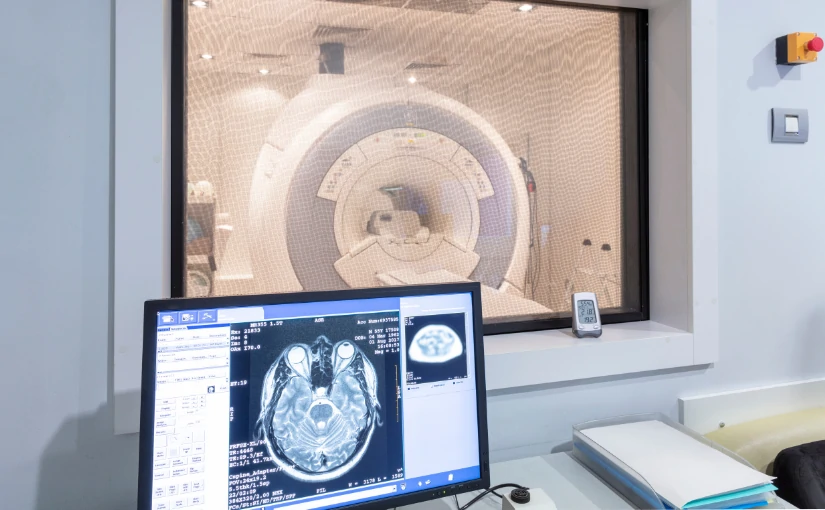
- Visualization and processingThe resulting image is displayed on a workstation, where different parameters can be adjusted. From modifying brightness, contrast and sharpness to adding annotations, measurements or labeling the image.
- Deletion of the plateAfter the process is completed, the plate is completely erased by intense light to remove any residual information. The process is then completed and the plate can be reused in another study.
Clinical workflow in computed radiography
The workflow in a computed radiography environment is systematic and designed to optimize time and ensure patient traceability. Although it is a more agile and efficient system than traditional development, it is not as immediate as direct digital radiology. Below are the different phases of the computed radiography workflow:
- Patient identification and study prescriptionIt starts with the loading of the patient's record in the RIS system (Radiology Information System), where the parameters of the request and the type of study required are defined.
- Image acquisitionThe technician positions the patient and makes the exposure with the CR plate in the cassette, as in a traditional X-ray.
- Digital reading of the cassetteAfter exposure, the cassette is transferred to the CR reader, where the latent image is digitized through the process described above.
- Processing and post-productionThe digital image is processed by specific software, allowing the technical parameters to be adjusted to optimize diagnostic visibility.
- Technical and medical validationThe technician checks the quality of the image before sending it to the radiologist, who will perform the clinical interpretation and generate the diagnostic report.
- Distribution and archiving: Finally, the image is stored in the PACS system (Picture Archiving and Communication System) and is included in the patient's electronic medical record.
Advantages of computed radiography
The adoption of computed radiography systems brings a number of important benefits to both healthcare personnel and medical facilities:
- Reduction in the use of chemicalsDoes not require the use of liquids or developer, which reduces environmental impact and biohazards.
- Reuse of platesPhosphor plates can be reused. Therefore, it offers great economic savings in the medium term.
- Improved image qualityCompared to analog radiology, CR offers better sharpness and digital adjustability.
- Easy integration into existing digital systemsIt can be connected to workstations such as the PACS system, the RIS system or DICOM printers, facilitating the exchange and management of medical information.
- Adaptability to existing equipment: Many installations of old X-rays Traditional or traditional systems can continue to be used with CR systems, which minimizes the initial costs of digitization.
Limitations compared to other techniques
Despite its advantages, computed radiography has certain limitations when compared to more advanced technologies, such as direct digital radiology (DR) systems:
- Increased processing timeThe technician must physically handle the cassette, which lengthens the time between exposure and image display.
- Increased operational burden for technical staffThe reading and handling of the cassettes involves additional steps that do not exist in the DR technique, where the image appears automatically.
- Slightly lower image qualityIn situations where maximum resolution and diagnostic accuracy is required, such as in fine lung studies or mammography, DR usually offers better results.
- Maintenance costs of CR readersAlthough CR technology is more affordable than DR, it requires a specific reader that involves maintenance, calibration and, in some cases, replacement of parts.
What are the differences between computed radiography (CR) and direct digital radiography (DR)?
| Features | Computed Radiography (CR) | Direct Digital Radiology (DR) |
|---|---|---|
| Image capture | Requires cassette with phosphor plate | Digital sensor integrated in the equipment |
| Image acquisition time | Slow (requires scanning of the cassette) | Immediate (real time image) |
| Equipment handling | Manual intervention of the technician for each study | Automated, requiring fewer steps |
| Image quality | Good, but inferior to DR | Excellent resolution and detail |
| Cost of implementation | Moderate, reuses traditional equipment | High, requires investment in advanced technology |
| Reuse of the detector | Yes, with erasable phosphor plates | Yes, with integrated digital sensors |
| Uses | Centers with progressive transition to the digital environment | High-demand, fast-flowing hospitals and clinics |
Main uses of computed radiography in clinical practice
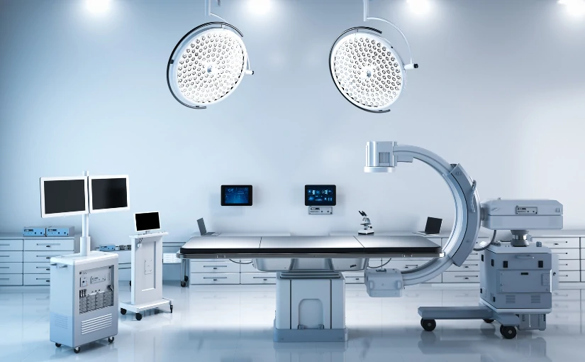
Computed radiography (CR) is used both in medical centers, hospitals and clinics as in mobile units. It offers a wide versatility, has a low operating cost and provides high compatibility with conventional equipment. These are its main applications in clinical practice:
General radiology
It is used for routine studies such as X-rays of the chest, abdomen, spine, pelvis and extremities. It is an ideal technique for initial and follow-up examinations.
Emergency and traumatology
In emergency departments, CR allows rapid imaging of fractures, dislocations or bone injuries without the need for chemical processing. It is very useful in the rapid evaluation of polytraumatized patients.
Postoperative control
It is used to verify the correct placement of prostheses, screws or osteosynthesis material after orthopedic surgery, as well as for the evolutionary follow-up of injuries.
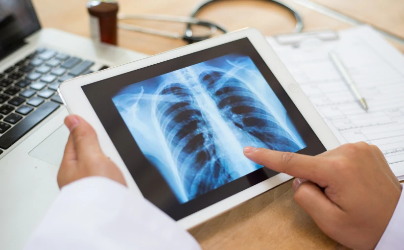
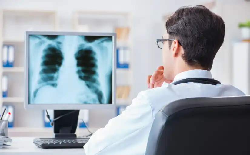
Thoracic and pulmonary evaluation
Chest radiography is one of the most frequent applications. It can detect infections, pleural effusions, nodules or signs of heart failure. CR facilitates digital contrast adjustment to improve the visualization of lung structures.
Dentistry and orthodontics
In some centers, computed dental radiography is used for orthopantomography, cephalometric studies or periapical radiographs, especially when compatible digital adapters are available.
Veterinary applications
Many veterinary centers use computed radiography as their primary imaging system because of its economy and ease of use, especially for radiographs of small and large animals.
Mobile units and health campaigns
Because of its portability and ease of installation, computed radiography is used in radiology trucks or mobile units.
Conclusion
Computed radiography is an effective, flexible and versatile medical technique. which has been key in the digitization process of diagnostic imaging services. It offers an efficient alternative for centers that wish to modernize without replacing all their equipment, adapting to multiple clinical environments.
Newer technologies, such as direct digital radiology, provide more automated and streamlined processes. Nevertheless, CR is still a viable alternative that can be used especially in small and medium-sized medical centers, mobile units or services with limited budgets that require a progressive transition to digital systems.
If your clinic needs advice on which computerized, conventional or direct radiography equipment is most suitable for your center, at 4D Médica we have the solution for each particular case. Request information without obligation.
Bibliography

CEO of 4D Médica. Expert in marketing and distribution of medical equipment.
![]()