Customer service: 910 020 252 4dmedica@4dmedica.com
How an ultrasound scanner works: Origin, process and clinical application

How an ultrasound scanner works: Origin, process and clinical application

At present, the ultrasound scanner has established itself as a fundamental tool in the area of diagnostic imaging. This device uses the ultrasound technology to obtain accurate, real-time images of the internal structures of the human body, facilitating the evaluation of organs, tissues and blood vessels. without the need for invasive procedures.

The ability of ultrasound to provide detailed, safe and rapid information has revolutionized clinical practice. The use of this medical equipment enables healthcare professionals to detect and monitor a wide variety of pathologies in an early and effective manner. In addition, its versatility and portability has extended its use to multiple medical specialties.

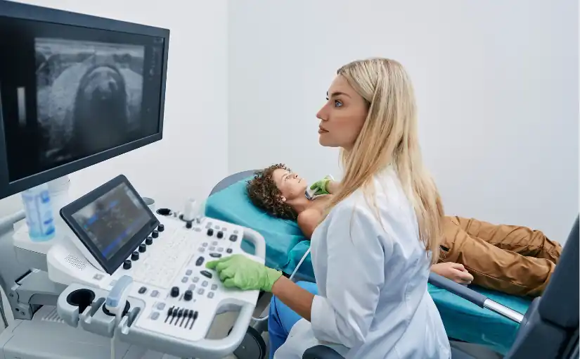
The use of the ultrasound scanner is used to perform ultrasounds in order to analyze organs and tissues internally. It is one of the most widely used medical techniques, as it is a fast, effective and non-invasive method. It is mainly used to detect diseases and anomalies, to monitor the health of patients, to study the development and growth of the baby during pregnancy, as well as to guide certain medical procedures.

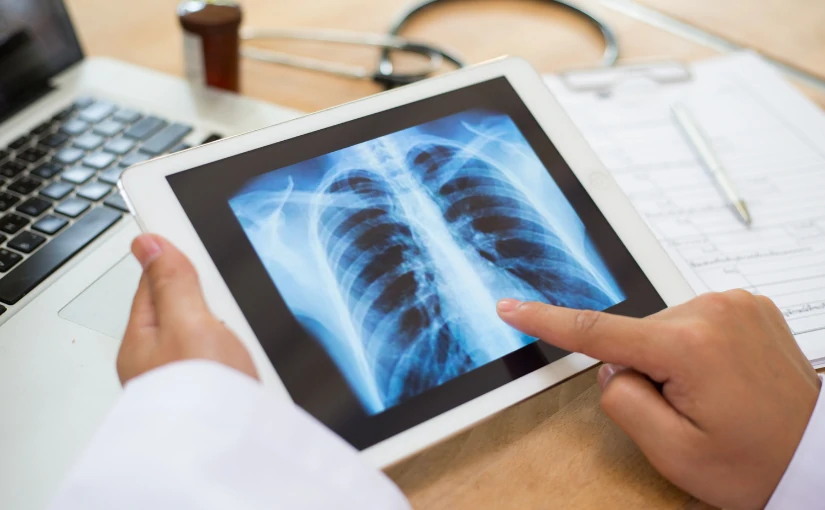
In contrast to other imaging techniques, such as X-rays or the Computed Axial Tomography (CT) scanultrasound does not use ionizing radiation, which makes it a safer technique. In addition, its portability and ease of use has enabled its integration in consultation rooms, emergency rooms and intensive care units, facilitating real-time clinical decision making and improving patient care.

In the following article, we analyze the origin of this medical equipment up to the present day, how an ultrasound scanner works, as well as its applications in clinical practice.

Origin of the ultrasound scanner: From its beginnings to the present day.

The development of the ultrasound scanner is closely linked to the development of ultrasound technology and its application in the medical field.

Early studies: Discovery of the piezoelectric effect

The first studies on ultrasonic waves date back to the end of the 19th century, when the French physicists Pierre and Jacques Curie discovered in 1880 the piezoelectric effect. This physical phenomenon consists of the ability of certain materials, such as quartz and some ceramic crystals, to generate an electrical charge when subjected to mechanical pressure.

The importance of the piezoelectric effect in ultrasound is fundamental, since it constitutes the basis of ultrasound transducer or probe operation. In practice, piezoelectric crystals located in the transducer convert electrical signals into ultrasonic vibrations (ultrasound waves), which are transmitted to the patient's body. Through the piezoelectric effect, these echoes are transformed into electrical signals that are processed by the ultrasound scanner to generate images in real time.

Development of the first ultrasound scanner: From the piezoelectric effect and ultrasound to the medical field

Following the discovery of the piezoelectric effect, the phenomenon was initially applied in industrial and military fields, particularly in the development of sonar devices for underwater detection during World War I and World War II.

However, the potential of ultrasound and the piezoelectric effect to generate and receive acoustic waves did not go unnoticed by the scientific and medical community. The adaptation of this technology to the medical field began in the middle of the 20th century. The Scottish physician Ian Donald, together with engineer Tom Brownwere the pioneering researchers who began to apply the piezoelectric effect to clinical explorationThe first of its kind, it laid the foundations of medical ultrasound.

Specifically, it was in the 1950s, when researchers developed the first clinical ultrasound prototype. Initially, ultrasound was used in obstetrics to visualize the fetus and detect pathologies during pregnancy, which was a revolution in prenatal monitoring.

From the 1960s and 1970s to the present day: Advances in ultrasonography

During the 1960s and 1970s, ultrasound technology made significant advances. It became from still images to real-time ultrasound scanswhich made it possible to observe the movement of internal organs and structures. Subsequently, the incorporation of the Doppler effect made possible the study of blood flow and vascular evaluation.The new ultrasound system has been designed to further expand the clinical applications of the ultrasound scanner.

In recent decades, the development of technology and information technology has made possible the emergence of more compact, portable ultrasound scanners with higher image resolution. Today, the ultrasound scanner is a safe, effective and versatile tool used in a wide variety of specialties, from emergency medicine to cardiology, gynecology and internal medicine. As a result, it has become an indispensable piece of medical equipment in medical practice.

Next-generation ultrasound scanners: innovation, technology and artificial intelligence

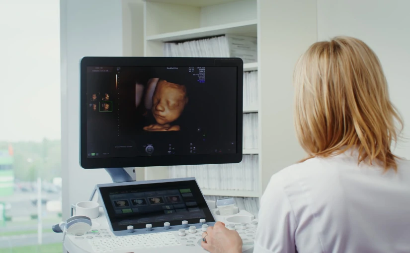
In recent years, technology has advanced greatly in the field of medicine. The state-of-the-art ultrasound scanners offer images in 3D, 4D and 5D technologyand therefore allow for visualize the inside of the human body in motion and in real time.

One of the most recent innovations is the ultrasound scanners that incorporate digital processing systems that apply the artificial intelligence in medical image analysis. The use of a AI software in ultrasound equipment provides greater speed, efficiency, safety and diagnostic accuracy, providing advanced and detailed analysis that improves clinical decision making.

In this area, it is worth mentioning the use of state-of-the-art ultrasound scanners to visualize the fetus in real time. It is known as emotional ultrasound and allows parents to get to know the baby before it is born. This type of ultrasound combines the 3D technologywhich provides three-dimensional images, with 4D and 5D technologyThe newborn's movements can be seen in real time with high image clarity and quality. With this, the baby's main movements can be seen. From yawning, opening the eyes and moving to changing position.

How an ultrasound scanner works: Step-by-step procedure

The ultrasound scanners are an essential tool in medical practice. Understand its operation and the workflow during an ultrasound examination is essential to ensure diagnostic quality and patient safety. Below, we discuss how an ultrasound scanner works and the step-by-step procedure:

1. Preparation of the patient and application of the conductive gel

First of all, the patient is instructed on the position The position to be used depends on the area to be scanned and the type of diagnosis to be made. Before starting the scan, the conductive gel on the patient's skin. It has the function of eliminating the air that is generated between the skin of the area to be examined and the transducer or ultrasound probe, facilitating the transmission of ultrasonic waves.

Selection of transducer type

One of the most important components of the ultrasound scanner is the transducer or probe. There are different types of transducersEach is designed to scan different regions and depths. While linear transducers are used for vascular and superficial studies, convex models are useful for deep abdominal scans.

Therefore, the medical professional will be in charge of selecting the type of transducer, connect it to the equipment and verify its correct operation. before starting the study.

3. Ultrasound emission and reception

Once the transducer is prepared, the operator places it on the gel-covered area. The transducer emits high-frequency ultrasound waves that penetrate the patient's internal tissues. When these waves pass through the body and are reflected at the interfaces of the different tissues and organs, the reflected waves, known as echoes, return to the transducer..

4. Echo pickup

The transducer also acts as a receiverby detecting the reflected waves (echoes) that are generated from the various internal structures. These echoes contain information on the location and characteristics of the traversed tissuesThis allows us to analyze the state and functioning of the different organs.

5. Image processing

Echoes picked up by the transducer are converted into electrical signals.These images are processed by the ultrasound console through different algorithms. The result is the generation of two-dimensional or three-dimensional images in real time that are displayed on the screen of the equipment.

By analyzing medical images, the operator is able to observe the anatomy and movement of internal organs and structures. In turn, with the use of the Doppler mode, the blood flow in the tissues can be studied.

6. Systematic exploration

The professional performs a methodical sweep by moving the transducer over the area of interest to be analyzedThe different sections (longitudinal, transversal, oblique) are obtained to fully examine the organs and structures. This systematic exploration is the key to obtain a complete and detailed diagnosisThe results of the study should be documented in a way that does not omit relevant findings, and the results should be adequately documented.

7. Adjustment of image parameters

During scanning, the operator can adjust various parametersThe display mode (2D, 3D, Doppler) can be set from the gain (brightness), depth and focus to the display mode (2D, 3D, Doppler). In this way, you can optimize image quality and adapt it to the anatomical characteristics of the patient.

8. Interpretation of medical images

After performing the examination, the physician is in charge of analyze the images obtained in real timeThe data can be used to identify possible alterations and to make static captures or recordings of relevant sequences. By means of these recordings, the final report can be fully documented, which will serve as the basis for the diagnosis and the clinical decision making.

9. Completion and cleaning

At the conclusion of the study, the operator removes the gel from the patient's skin. Subsequently, a protocol for disinfection and cleaning of the equipment usedThe transducer and the contact surface between each patient.

This structured process makes ultrasound a fast, safe, non-invasive and highly versatile technique, facilitating the accurate assessment of multiple organs and pathologies in daily clinical practice.

Main clinical applications of ultrasound

The use of ultrasound scanners covers practically all medical specialties. The main clinical applications include the following areas:

Obstetrics and gynecology

Ultrasound is essential in the pregnancy monitoringIt is used to evaluate fetal development, the location and viability of the pregnancy, the detection of congenital anomalies and the control of obstetric complications. It is also used for the study of gynecological pathologiesas ovarian cysts, uterine myomas or endometrial alterations.

Cardiology

The echocardiogram is an essential technique for the assessment of cardiac anatomy and functionallowing to diagnose valvular diseases, cardiomyopathies, heart failure, congenital heart diseases and evaluate blood flow using the Doppler mode.

Internal medicine and gastroenterology

The abdominal ultrasound allows examination of organs such as the liver, gallbladder, pancreas, kidneys, spleen and bladder. It therefore plays a key role in the diagnosis of masses, cysts, stones, inflammations and other pathologies. It is also used in the assessment of ascites and in the control of interventional procedures.

Vascular exploration

Through the Doppler ultrasoundthe blood flow in arteries and veinsIt is therefore used in the diagnosis of deep vein thrombosis, venous insufficiency, arterial stenosis, aneurysms and other vascular diseases.

Musculoskeletal

Allows you to study muscles, tendons, ligaments, joints and soft parts, facilitating the diagnosis of sports injuries, tears, tendinitis, bursitis, hemorrhages and subcutaneous masses.

Urology

It is used for assess the prostate, bladder, testicles and kidneysbeing useful in the diagnosis of prostatic hyperplasia, lithiasis, tumors and other urological alterations.

Pediatrics

Ultrasonography is especially useful in the study of pediatric pathologies, such as the hip dysplasia, hydrocephalus, renal malformations and abdominal alterations in newborns and infants.

Guidance on interventional procedures

The ultrasound scanner facilitates the performance of biopsies, drainages, punctures, catheter placement and other interventionsThe procedure is safer and more accurate.

Emergency medicine and intensive care

Its speed and portability allow immediate diagnosis of serious pathologies such as pleural effusions, hemoperitoneum, pneumothorax, cardiac tamponade and rapid assessment in polytraumatized patients (FAST ultrasound).


Conclusion

Ultrasound has established itself as an essential tool in clinical practice.It offers an accurate, efficient, safe and real-time medical analysis of the different internal organs and tissues. Its non-invasivenessThe absence of ionizing radiation and its versatility to be adapted to multiple specialties make it an indispensable resource both in the initial evaluation and in the follow-up of numerous pathologies.

Your portability and speed facilitate clinical decision making in a variety of settings, from outpatient to emergency situations. From its origins to the present day, the use of ultrasound has revolutionized medical practice by improving the quality of health care and making a decisive contribution to a early, accurate and safe diagnosis for patients.

If you want to obtain more information about ultrasound scanners or other medical diagnostic equipment, you can contact us. Our 4D team will give you advice to find the best solution for your clinic or hospital.

Contact 4D


Bibliography

García, J., & González, A. (2007). Ultrasound methodology and techniques. Physical principles and image formation. Medicina de Familia SEMERGEN, 33(2), 83-92. Retrieved May 20, 2025, from. https://www.elsevier.es/es-revista-medicina-familia-semergen-40-articulo-metodologia-tecnicas-ecografia-principios-fisicos-13109445

Spanish Society of Pediatric Intensive Care (SECIP) (2018). Basic fundamentals of ultrasound. Retrieved May 20, 2025, from. https://secip.com/images/uploads/2018/09/1-FUNDAMENTOS-BASICOS-DE-ECOGRAF%C3%8DA.pdf

Authorea (n.d.). Ultrasound: Physical principles and clinical applications. Authorea. Retrieved May 20, 2025, from. https://www.authorea.com/doi/full/10.22541/au.172660489.98960333

Luis Daniel Fernandez Perez

Director of Diagximag. Distributor of medical imaging equipment and solutions.

Telemedicine: What it is, how it works and its relationship to AI

Telemedicine: What it is, how it works and its relationship to AI

In recent decades, the introduction of new technologies in the health sector has enabled the development of new forms of healthcare. It is becoming increasingly common for patients to receive a remote medical care by healthcare professionals, through video consultations, remote monitoring or digital diagnosis. This is what is known as telemedicine.

The main objective of the incorporation of new Information and Communication Technologies (ICT) in medicine was to bringing health services closer to the population who resided in remote regions and who had a reduced health accessibility and resources. Over the years and with the development of new technological innovations, including the emergence of artificial intelligence (AI), ICT became a key tool in the development to improve the quality and efficiency of healthcare.

What is it and how did it come about? In the following article, we explain what telemedicine is, how it works and its relationship with AI, as well as its different advantages and disadvantages.

What is telemedicine?

Telemedicine is the a set of medical practices that use information and communication technologies (ICT) to provide remote health care services.. This includes medical consultations, diagnoses, treatments, clinical follow-up, issuing prescriptions and preventive guidance, without the need for the patient and the professional to be physically present in the same place. For this purpose, telemedicine allows access to health services through electronic devices such as computers, tablets or smartphones, through secure digital platforms.

The World Health Organization (WHO) defines it as: "The provision of health services at a distance through the use of information and communication technologies for diagnosis, treatment, disease prevention and research."

Telemedicine and eHealth (digital health): What are their differences?

At the same time, the eHealth (digital health) conceptThe term "health informatics", which encompasses a broader term and is located between medical informatics, public health and commercial interest. It refers to the use of information and communication technologies (ICT) to improve, support and optimize the delivery of healthcare services and the management of the healthcare system.

Although often used synonymously, eHealth and telemedicine are not the same thing, although they are closely related. While eHealth telemedicine connects physicians and patients at a distance, eHealth encompasses all types of digital tools that improve health.. From an app to a hospital management system, such as the PACS system or the RIS system.

In this context, the telemedicine concept is part of the eHealth ecosystem. and focuses specifically on the provision of medical services, providing remote consultation, diagnosis and treatment.

Origin: Technological stages and evolution

Telemedicine is not a recent concept. Its origin dates back to the 1950s.The first remote communication systems began to be used for medical purposes. One of the first documented cases was in the United States, where a telephone line was used to transmit radiological images between hospitals. The following stages can be distinguished:

Early decades: 50's-70's

In the late 1960s, the NASA played a key role in the development of telemedicine. Faced with the need to be able to monitoring the health of astronauts during space missionsThe development of technologies capable of recording and transmitting biometric data remotely was promoted. These advances were also applied in rural Alaska through the STARPAHC (Space Technology Applied to Rural Papago Advanced Health Care) program, considered one of the first structured telemedicine projects.

Technological evolution: 1980s to 2000

During the 1980s and 1990s, the improvement of computer technology and satellite communications allowed the expansion of telemedicine services, especially in rural and military environments. The use of telemedicine systems for teleconsultation, remote diagnosis and exchange of clinical information between hospitals.

In these years, video calls started to be used for dermatology, psychiatry and radiology consultationsThe use of telemedicine was limited, however, by the high cost of equipment, lack of infrastructure and poor digitization of medical records. However, the use of telemedicine was limited by the high cost of equipment, the lack of infrastructure and the scarce digitalization of medical records.

Digitalization and the rise of the Internet: Years 2000-2010

With the the advent of the Internet, personal computers and smartphones.telemedicine took a qualitative leap forward. Starting in the 2000s, telemedicine began to develop more accessible platforms for online consultations, chronic patient follow-up, diagnostic test referrals and distance medical education. The first electronic medical record systems were also integrated, which facilitated networking among professionals from different health centers.

Telemedicine today

Although telemedicine was already in existence, the COVID-19 pandemic in 2020 marked a definitive turning point in its adoption worldwide. Faced with the need to avoid travel and minimize the risk of contagion, many healthcare systems began to offer video-call consultations, electronic prescriptions, remote monitoring and online psychological care. Today, telemedicine has established itself as a standard medical solution in many countries and has been integrated into public and private healthcare services.

The relationship between telemedicine and artificial intelligence

The development of the artificial intelligence in medicineIn recent years, remote monitoring devices and predictive algorithms are driving the rise of telemedicine. In recent years, the combination of telemedicine and artificial intelligence is transforming the way medical care is delivered to patients. Both technologies, when combined, improve the efficiency, accuracy and accessibility of the different healthcare services. However, although they are two complementary tools, each one has its own operation and its own specific characteristics. medical applications specific.

On the one hand, the telemedicine enables health care professionals to to care for patients remotely. On the other hand, the IA is responsible for analyze large volumes of medical data, detect patterns, automate tasks and suggest diagnoses or treatments.

Therefore, when used together, more agile, intelligent and personalized systems are created. At present, the use of AI software to improve the efficiency and accuracy of medical diagnosis, as well as facilitate patient management and healthcare.

Types of telemedicine: We analyze how it works and what it is used for.

Since the emergence of telemedicine and with the evolution of different technologies, different types of telemedicine have been developed that define the concept as we know it today. Below, we analyze how each of these modalities works and what they are used for:

Teleconsultation

The medical consultation represents the basis of clinical practice in the field of medicine. For this reason, teleconsultation is the most commonly used modality. It is based on the search for local or external medical information or medical advice, using information and communication technologies.

Communication between the patient and the healthcare professional can be done directly or through third parties. Thus, there are two different types of teleconsultation: asynchronous and synchronous.

  • Asynchronous teleconsulting

In this type of teleconsultation, known as asynchronous, medical care is provided by means of the The physician will send clinical information and, subsequently, the physician performs the assessment and counseling.

Main advantages
  1. The parties involved do not have to be present at the transfer of information.
  2. It offers the ability to capture and store still and moving images of the patient, as well as audio and text, providing the healthcare professional with more clinical information.
  3. It is an economical and accessible modality, since it supports a large volume of work and analysis of medical tests.
  • Synchronous teleconsultation

Synchronous teleconsultation is developed in real timeand, therefore involves the participation of patients and health care professionals in sending information through the use of telecommunication technologies.

In this modality, videoconferencing stands out as the most commonly used technologyIt provides both visual and auditory contact with the patient. This facilitates pattern recognition and greater accuracy in making a medical diagnosis.

Main advantages
  1. Fast and effective diagnosis
  2. Better understanding between patients and professionals health care
  3. Integration of additional techniques that increase reliability
    of clinical information. This is the case of digital auscultation.
Main disadvantages
  1. Its execution involves a number of huge costs economically, since it requires a certain telecommunication infrastructure.
  2. Requires a increased demands on the time of medical professionals, as they must allocate time for the teleconsultation and, additionally, carry out a pre- and post-evaluation.

Teleeducation

It is defined as the use of information and telecommunication technologies for distance medical education practice. In this field, Internet technologies and videoconferencing are the means most commonly used by health professionals to increase their skills and put their knowledge into practice. Within tele-education, different modalities can be distinguished, depending on the way in which the information is transmitted:

  • Tele-education through teleconsulting

A physician who is an expert in a particular specialty provides a diagnosis to the query raised by a non-expert physicianintern or resident.

  • Clinical education via the Internet

Allows the access to various databases with medical and clinical articles and books. These include MedLine, Cochrane, the National Library of Medicine in the United States and the National Electronic Health Library in the United Kingdom.

  • Academic studies via Internet

Different universities, both public and private, offer courses and tele-educational programs, as well as virtual internships., where participants are evaluated and graded to obtain a set of competencies that will allow them to develop their professional career in the health area.

  • Public education through telemedicine

Refers to the medical education and communication offered on different topics related to public health. From diet, exercise and hygiene websites to different diseases, such as cancer and AIDS.

Telemonitoring

Telemonitoring is the use of information and communication technologies to obtain information regarding the patient's condition and status to determine if adjustments or changes to the proposed treatments are necessary.

This type of telemedicine allows professionals to monitor different aspects: physiological variables, test results, as well as images and sounds. It is usually performed at the patient's home or in nursing centers, which reduces costs and resources for the healthcare system.

Telesurgery

Telesurgery is based on the development and execution of surgeries where the surgeon acts by means of remote visualization and manipulation using electronic devices and high technology in telecommunications. The main objective of telesurgery is to offer surgical services to patients who, for reasons of inaccessibility, cannot be attended in person in hospitals and medical clinics. Telesurgery has two different modalities, which are discussed below:

  • Tele-surgery through tele-education or telementoring

Telesurgery by means of tele-education or telementoring is an advanced form of remote surgical training and medical care that combines telecommunications technology, real-time surgery and medical teaching techniques.

The telesurgery by means of telementoring is that a skilled surgeon (mentor) provides technical advice, corrections, instructions or live training during a surgical procedure being performed by a less experienced surgeon (trainee)even if they are in different geographical locations. This is achieved by means of videoconferencing systems, augmented reality, laparoscopic cameras or interactive platforms.

For its part, the surgical tele-education goes beyond the operating room and encompasses also theory sessions, case review, virtual classes and guided surgical simulationall from a distance.

  • Telepresential surgery

Telepresential surgery is an advanced modality of technology-assisted surgery that enables a surgeon to control surgical instruments remotely using robotic systems connected by high-speed telecommunications networks.

This is a form of telesurgery and allows the professional to act as if he were in the operating room through the use of technology. For example, the use of robotic arms, micro cameras and high resolution optical instruments.

Advantages and disadvantages of telemedicine

Advantages of telemedicine Disadvantages of telemedicine
Facilitates access to medical care from any location Does not allow complete physical examinations
Reduces travel and waiting times Dependence on devices and good internet connection
Saves costs for patients and healthcare facilities Digital barriers may exist for the elderly or people with few resources
Improved follow-up of chronic patients Some specialties are not compatible (e.g. surgery, dentistry).
Promotes continuity of care and preventive care Loss of human and nonverbal contact in the doctor-patient relationship.
Contributes to sustainability by reducing carbon footprint Medical data security and privacy risks

Main advantages of telemedicine

Telemedicine and technological innovations in the healthcare field have provided a number of benefits, driving the improvement of healthcare and medical diagnosis.

Access to health care from anywhere

Telemedicine makes it easier to people living in rural areas, with limited health care resources or with reduced mobility can receive medical care without the need to travel. In this way, it is characterized by bringing medical care closer to different groups. From the elderly and migrant population to patients with disabilities, which improves health equity.

Time savings and convenience

Avoids trips to health centers or hospitals and eliminates waiting times in consultation rooms. The patient can perform consultations from home, obtaining greater time flexibility and without interrupting his or her daily routine. On the other hand, consultations are shorter and more directThe work of healthcare professionals is optimized.

Cost reduction

It reduces costs for both patients and health centers. On the one hand, patients avoid the expenses associated with transportation, permit applications labor andIn many cases, it is also reduces the cost of consultation. And, for health centers, it represents a significant saving by reducing the need for physical infrastructure, logistical resources and on-site personnel. Thus, it favors the efficiency of the public and private system.

Effective chronic disease monitoring

Allows you to real-time patient monitoring with pathologies such as diabetes or hypertension, avoiding complications and improving adherence to treatment.

Better doctor-patient communication

Promotes a closer and continuous careThe system is ideal for resolving doubts, reviewing tests or following up with the patient without having to visit the health center in person.

Optimization of the healthcare system

Reduces the burden on emergency and primary care by filtering non-urgent consultations and improving medical resource management.

Positive impact on the environment

By reducing the number of trips, we also carbon footprint is reduced associated with medical care.

Boosting digital health and education

Allows to integrate educational content and interactive resources to assist the patient to take care of your health from home. And, at the same time, it makes the medical training for professionals through tele-education.

Greater control and security

Digital platforms protect the patient privacy and generate a electronic medical record with better traceability and follow-up. In this way, patients can consult their records, tests and treatments digitally.

Disadvantages and limitations of telemedicine

Although telemedicine offers numerous benefits, it also presents a number of limitations and challenges:

Direct physical examinations are not possible

The main limitation is that direct physical examinations are not permitted, which can make diagnosis difficult in complex or urgent cases. Some diseases require palpation, auscultation or immediate tests that can only be done through a face-to-face consultation.

Technology dependence and digital divide

For telemedicine to work properly, it is necessary to have a good Internet connection, the use of appropriate medical devices and knowledge of the use of digital applications. Not everyone has access to digital tools or the Internet. This may exclude older people or those with limited technological resources.

Difficulties in the patient-physician relationship

Physical contact and nonverbal communication are key in the clinical relationship. In some cases, telemedicine may lead to a feeling of distance or lack of empathy between health professionals and patientsespecially in consultations with sensitive diagnoses.

Data privacy and security risks

The use of digital platforms entails risks of leakage or misuse of personal and medical information if the following guidelines are not applied. appropriate cybersecurity measures.

Technical limitations and connection failures

Technical problems such as network outages, poor image or sound quality, as well as software malfunctions can interrupt or hinder consultations, affecting the quality of service.

Restrictions on certain medical specialties

Not all areas of medicine are well suited to the virtual environment. For example, surgery, traumatology or dentistry require mandatory physical presence, and telemedicine can only complement some processes, not replace them.

Telemedicine represents the combination of technology and health services. Over the years, its evolution has driven an increasingly complete and efficient healthcare system. In this context, the relationship between telemedicine and artificial intelligence stands out, since it offers a higher quality of health careThe aim is to provide a more accurate medical diagnosis and the development of medical treatments customized to the real needs of patients.

Bibliography

Otero López, M. J. (2012). Telemedicine: A tool also in the rural environment. Primary Care, 44(10), 574-575. https://doi.org/10.1016/j.aprim.2012.03.016

Torres, M. R. R. R., & Collado, M. E. M. (2016). Telemedicine: Current status and perspectives. Clínica Las Condes Medical Journal, 27(5), 571-577. https://doi.org/10.1016/j.rmclc.2016.09.003

Peña González, A., & Córdova Alcaraz, L. (2017). Application of telemedicine in primary health care. Cuban Journal of Information in Health Sciences (ACIMED), 28(2), 135-145. https://www.redalyc.org/pdf/2611/261120984009.pdf

Sánchez-Guzmán, M. A., & González, S. M. (2015). Telemedicine: technological innovation in health. Iberoamerican Journal of Educational Research and Development, 6(12), 1-16. https://www.redalyc.org/pdf/2310/231019866002.pdf

Luis Daniel Fernandez Perez

Director of Diagximag. Distributor of medical imaging equipment and solutions.

Classification of medical equipment according to risk

Classification of medical equipment according to risk

In the healthcare sector, patient safety comes first. For this reason, medical equipment must comply with certain regulations to ensure its reliability, efficiency and traceability. The classification of medical equipment regulatory requirements can be tailored according to the level of risk represented by each medical equipment for patients.

Depending on the type of riska series of controls and evaluations that must be carried out before they reach the market and can be marketed.. The higher the risk, the more rigorous and demanding the processes of clinical evaluation, quality control, technical documentation and post-marketing follow-up become. In the following article, we discuss how medical products are classified. in accordance with MDR (Medical Device Regulation).

Entry into force of the MDR standard for the classification of medical equipment

The MDR (Medical Device Regulation) regulation, officially known as the Regulation (EU) 2017/745 on medical devicesis the legal framework in force in the European Union for the regulation of medical devices. It is a European regulation that replaces the former Directive 93/42/EEC (MDD) and Directive 90/385/EEC on active implantable devices. In contrast to the directives, the MDR has a direct effect in all EU Member Stateswithout the need to adapt national laws.

It was approved on April 5, 2017 with the aim of reinforcing the safety, traceability and efficacy of these products on the European market, but its official entry into force did not arrive until May 25, 2017. As of May 26, 2021 and after a transition period of 4 years, its application became mandatory throughout the European Union.. For certain products certified under the old MDD standard, there is an extended transition period until 2027-2028.

Main changes in MDR regulations

The MDR replaced Directive 93/42/EEC (MDD). with a more precise and strict regulation. This regulation established new more detailed rules for classifying devices according to risk level. To this end, a number of specific criteria were applied, such as duration of use, invasiveness, area of the body affected and type of operation (active or passive). At the same time, it also added specific rules for medical software and the AI softwarewhich were not sufficiently covered before.

The main changes introduced were as follows:

  • More specific and strict classification rules, strengthening clinical and technical evaluation.
  • Increased control and new classification of implantable products and medical software
  • More rigorous assessment by notified bodies
  • Enhanced post-marketing surveillance requirements
  • Introduction of EUDAMED system for increased traceability and transparency
  • Direct and uniform application in all EU countries
  • Adapting the regulatory framework to new technologies, such as medical software and artificial intelligence applied to medicine

Classification of medical equipment according to MDR

The MDR regulation establishes a classification system in four different classes (I, IIa, IIb and III) according to the potential risk that the medical equipment represents for the user.

Class Risk Features Examples
Class I Under
  • Non-invasive
  • External or superficial use
  • No critical function
  • Bandages
  • Gloves
  • Simple thermometers
Class IIa Moderate
  • Short-term invasive
  • They may have software
  • Limited interaction with internal organs
  • Headphones
  • Short catheters
  • Basic medical software
Class IIb High
  • Invasive of medium/long duration
  • Act on vital functions
  • Prolonged use in internal organs
  • Respirators
  • Infusion pumps
  • Neonatal incubators
Class III Very high
  • Long-term implantable
  • Affect vital functions
  • Use in circulatory or nervous system
  • Pacemaker
  • Stents
  • Therapeutic AI software


Class I - Low risk

Class 1 medical devices are non-invasive equipment, for temporary or external usewhich do not interact directly with physiological functions critical body parts. Their design and use involves a minimal risk for the patient.

Main features

  • They do not require electricity or software to operate, they are "passive".
  • Used on the surface of the body or in a superficial manner
  • They may include variants such as:
    • Is (sterile)
    • Im (measuring function)
    • Go to (surgical reusables)

Examples

  • Gauze, bandages and sticks
  • Mercury-free thermometers
  • Non-sterile medical gloves
  • Manual wheelchairs

Type of evaluation

Generally, it is requires manufacturer's self-certificationexcept for the variants Is, Im and Ir, which require assessment by a notified body.

Class II - Moderate and high risk

Class 2 medical devices include two different modalities: Class IIa devices, which have a moderate risk, and Class IIb devices, which have a high risk.

Class IIa - Moderate risk

Includes short-term invasive medical deviceswhich are in use for less than 30 days, or active, which may have a moderate impact on health of the patient. This type of medical products can enter body cavities o use in non-critical diagnostic or therapeutic procedures.

Main features
  • Invasive through natural orifices or with limited medical intervention
  • Can be electrically powered or contain software
  • The risk is higher than in Class I, but still limited.
Examples
  • Hypodermic needles
  • Short-term catheters
  • Headphones
  • Non-critical monitoring software
Type of evaluation

Requires the participation of a notified body which evaluates technical documentation and clinical evidence, although it is less complex than in the higher classes.

Class IIb - High risk

It includes devices that can have a significant impact on vital physiological functions, which are long-term invasive or acting on critical internal organs. Also included is the software that directly influences relevant clinical decisions.

Main features
  • Invasives of medium or long duration
  • They act on the circulatory system or the central nervous system (if not for prolonged use)
  • Includes devices that deliver automatic treatments
Examples
  • Respirators
  • Neonatal incubators
  • Hemodialysis equipment
  • Diagnostic imaging software with AI
  • Programmable infusion pumps
Type of evaluation

Requires comprehensive clinical evaluation, technical review by a notified body and strict compliance with regulatory requirements.

Class III - Very high risk

Class 3 devices present the highest level of risk, since they may have a direct impact on vital functions or its use may involve critical intervention in the human body. Includes permanent implantable devices and stand-alone software for diagnosis or therapy.

Main features

  • Long-term or permanent implants
  • Long-term invasive devices in the central nervous system or circulatory system
  • Software with autonomous therapeutic functions

Examples

  • Pacemaker
  • Intracoronary stents
  • Cardiac valve prostheses
  • Brain implants
  • Artificial intelligence software provides oncology treatment solutions

Type of evaluation

Requires a mandatory full clinical evaluationincluding studies with patients. To this end, the notified body is involved at each stageThe following steps are required: development, manufacturing, documentation, post-sales surveillance. This type of medical equipment, being so high-risk, requires intensive post-marketing follow-up.

Factors that determine the classification of medical devices according to the MDR

The MDR regulation (Regulation (EU) 2017/745) establishes specific criteria for classifying medical devices according to their level of risk to the patient and the healthcare professional. What are the factors that determine the classification according to their risk?

Duration of use

This refers to the length of time the device remains in contact with the body. The longer the duration of contact, the greater the potential risk.

  • Temporary useLess than 60 minutes
  • Short-term useBetween 60 minutes and 30 days
  • Long-term use: More than 30 days

Degree of invasiveness

Evaluates whether and how the device penetrates the body. Implantable or surgical devices are rated higher.

  • Non-invasiveDoes not penetrate the body (e.g. bandages, external thermometers).
  • Invasive through natural orificesIt enters through the mouth, nose, ear, urethra, etc.
  • Surgical invasiveRequires medical intervention for insertion
  • ImplantableRemains inside the body for a prolonged period of time.

Affected body part

The MDR standard checks the site where the device acts in order to assess its risk. This risk increases when it affects a critical area of the human body.

  • Body surface or skin: lower risk
  • Internal organs or sterile cavitiesintermediate risk
  • Central nervous system or circulatory system: high risk

Active or passive use

Active devices can fail and their impact on the organism is greater, so they tend to be classified in higher classes.

  • Passive deviceOperates without energy source (e.g., syringes, dressings).
  • Active deviceRequires electrical or mechanical power to operate (e.g., respirators, infusion pumps).

Medical purpose

Another aspect that should be analyzed is the function performed by the device in the medical treatment or diagnosis. The higher the functional complexity and clinical relevance, the higher the risk in the classification. In this context, the following medical purposes can be differentiated:

  • Basic monitoring
  • Diagnose, treat or monitor medical conditions
  • Supports physiological functions
  • It is used for prevent diseases
  • Directly influences clinical decisions

Use of software

The MDR establishes clear rules for classifying medical software according to its use and clinical applications. The risk does not depend on the hardware, but on the purpose and clinical impact of the software.

  • Data management softwareClass I: Included in Class I
  • Software that aids in diagnostics or clinical decisionsClass IIa or IIb: Incorporated in Class IIa or IIb.
  • Autonomous software that makes therapeutic decisionsClass III: They are included in Class III because of increased risk.

Nature of the content covered

It is important to analyze whether the devices come into contact with the human body or alter the chemical composition of the organism. What options can we find depending on the nature of the content?

  • The device enters contact with blood, body fluids, or tissues
  • Modifies substances (chemically or thermally)
  • Administers medication or energy

The MDR regulation comprehensively analyzes how, where, how long and for what purpose a medical device is used. Each of these factors contributes to assigning it a risk class (I, IIa, IIb or III), which determines the legal and clinical requirements necessary to market it.

Importance of proper hazard classification of medical equipment

Classifying medical devices according to their risk is essential to ensure the safety of patients and users, and also to ensure that products comply with the appropriate regulatory requirements before they are marketed or used. What is the role of proper classification in the healthcare sector?

Protection of patients' health and lives

The classification makes it possible to identify the potential hazard level of a device. In this way, the necessary controls can be put in place to prevent failures that could cause harm to patients or healthcare professionals.

Determines the level of regulation and control

Higher-risk devices (Class III) require more rigorous clinical evaluations, testing, certification and post-market surveillance. In contrast, low-risk (Class I) devices follow simpler procedures, such as self-declaration of conformity by the manufacturer. This ensures that each device goes through a process commensurate with its level of risk.

Guidance to manufacturers and developers

Another of its functions is to assist manufacturers in understand key technical, clinical and documentary requirements that must be complied with according to the class of the device. Assessments and controls according to the risk of the medical equipment allow planning the process of development, validation, registration and market launch in an efficient and legally compliant manner..

Facilitates the work of health authorities

Regulatory authorities may prioritizing inspections and audits according to the risk associated with the product. This simplifies decision-making to authorize or restrict the use of certain devices.

Establishes trust in the marketplace and among users

Healthcare professionals and patients can be confident that they can rely on a product has been evaluated proportionally to the potential risks it represents. In this way, the promotes transparency, traceability and efficient management of incidents or product recalls.

It is a mandatory legal requirement

In most countries, classifying medical devices according to their risk is a legal requirement for approval and marketing (as in the European MDR Regulation, the FDA in the USA or the Chilean Institute of Public Health).

The classification of medical devices according to their risk is not only a regulatory procedure, but an essential tool to protect the health of patients and professionals, guarantee quality and make the entire healthcare system more efficient. If you work in the medical, technological or regulatory sector, knowing and applying this classification is the first step to ensure that your products reach the market in a safe, legal and responsible way.

Bibliography

Eurofins (n.?f.). What is a medical device/medical device? Eurofins Spain. Retrieved March 27, 2025, from https://www.eurofins.es/consumer-product-testing/industrias/productos-sanitarios/que-es-un-dispositivo-medico-producto-sanitario/

DQS Global (n.?f.). Understand the classification of medical devices according to the EU Medical Devices Regulation. Retrieved March 27, 2025, from https://www.dqsglobal.com/es-es/formacion/blog/comprender-la-clasificacion-de-los-productos-sanitarios-con-arreglo-al-reglamento-sobre-productos-sanitarios-de-la-ue

European Commission (2021). Medical Devices - Sector. European Commission - Public Health. Retrieved March 27, 2025, from https://health.ec.europa.eu/medical-devices-sector/overview_en

Public Health Institute of Chile (2019). Medical Device Hazard Classification Guidance. Retrieved March 27, 2025, from https://www.ispch.cl/sites/default/files/Guia_de_Clasificacion_de_Dispositivos_Medicos_Segun_riesgo_Formato_Institucional.pdf

Spanish Agency of Medicines and Health Products (s.?f.). Medical devices. Government of Spain - AEMPS. Retrieved March 27, 2025, from https://www.aemps.gob.es/productos-sanitarios/

European Union (2017). Regulation (EU) 2017/745 of the European Parliament and of the Council of 5 April 2017 on medical devices.. Official Journal of the European Union. Retrieved from https://eur-lex.europa.eu/legal-content/EN/TXT/?uri=CELEX%3A32017R0745

Luis Daniel Fernandez Perez

Director of Diagximag. Distributor of medical imaging equipment and solutions.

Emotional ultrasound: Getting to know the baby before birth is possible

Emotional ultrasound: Getting to know the baby before birth is possible

Can you imagine being able to see the baby's gestures during pregnancy? From how it moves and yawns, the moments when it opens its eyes, when it changes position and even how it plays with the umbilical cord. Knowing the baby before it is born and see all their real-time movements is possible through emotional ultrasound.

This is a type of ultrasound scan that goes from the beyond medical diagnosisoffering a more human and closer experience in the area of image diagnosis. Not only does it provide medical information, but it also plays a key role in strengthening the emotional bond between the parents and the baby during pregnancy and gestation.

Emotional ultrasound is one of the most innovative techniques in the field of ultrasound and, by means of ultrasound, the baby can be visualized in detail. The emotional ultrasound equipment that are used combine 3D technologywhich offers three-dimensional images, and 4D and 5D technology, which incorporates the movement of the fetus in real time with high image sharpness and quality. Therefore, emotional ultrasound not only ensures that the baby can be observed with a high resolution, but also that its gestures and movements can be seen in the mother's uterus.

In the following article, we discuss its main characteristics and differences with follow-up medical ultrasound scans, as well as all the advantages it offers.

Main features and advantages of emotional ultrasound

Emotional ultrasound provides added value by offering a more accurate closer experience of parents and family members with the babyWhat are its main characteristics and differences with a traditional medical ultrasound?

High image quality

Emotional ultrasounds use advanced technology that allows visualization of the fetus with high detail, generating sharp, moving images in real time.

Emotional bonding with the baby

The main objective of the ultrasounds is to check the correct development of the baby. However, the emotional ultrasound is a non-invasive test that provides a experience that is closer, more human and real for parents and family members. It is performed in a relaxing environmentThe nursery is equipped with soft music and adapted lighting to create a cozy environment. In some cases, family members are allowed to be present to share the moment and to be able to see the baby's movements and gestures.

In turn, the professionals who perform the ultrasound scan employ a warmer and closer approachexplaining every detail with sensitivity and empathy.

Complete monitoring of the fetus during pregnancy

In addition to visualizing the development of the baby in real time, emotional ultrasound is also used to perform a complete monitoring of the fetus during gestation. It is essential to analyze its neurophysiological statusas well as to detect possible anomalies and malformations.

Does not replace diagnostic medical ultrasound scans

It is important to keep in mind that, during pregnancy, follow-up medical ultrasounds should be performed. The emotional ultrasound does not replace in any case the different ultrasounds that the mother should have to evaluate the correct development and growth of the baby in the different stages of gestation. What are the different follow-up medical ultrasounds and when are they performed? We can distinguish the following:

  • Pregnancy confirmation ultrasound (week 6 and 8)It is developed to verify pregnancy and check that the embryo is in the mother's uterus.
  • First semester ultrasound scans (week 11 and 14)It is used to measure the length of the fetus, estimate the due date and check whether it is a single or multiple pregnancy.
  • Morphological ultrasound (week 18 and 22)It allows to examine in detail the organs and structures of the fetus. It helps to detect congenital malformations, evaluate natal growth and determine the sex of the baby.
  • Third trimester ultrasound (week 28 and 32)It is used to evaluate the baby's growth.
  • Prepartum ultrasound (36th and 40th week)This is the last ultrasound and is essential to check the baby's position, examine the approximate weight of the baby and the condition of the mother's amniotic fluid and placenta.

Use of ultrasound scanners with advanced technology

To perform the emotional ultrasound, specific medical equipment is used that incorporates the latest technology to visualize the baby in detail, with high image quality, sharpness and in real time. For this purpose, the following are used ultrasound scanners with advanced 4D and 5D technology.

Creation of memories in digital format

Emotional ultrasound not only offers real-time visualization of the baby, but also provides parents and family members to have a memory of that beautiful moment. The clinics that perform this type of 5D ultrasound provide the experience together with the delivery of the different ultrasound images and videos in digital format. so that parents can preserve, share and remember this beautiful memory of pregnancy and gestation.

When is it recommended to perform an emotional ultrasound?

Emotional ultrasound can be performed at any time during pregnancy. However, the most indicated time is between 25 and 30 weeks.since the baby is more developed and the baby's movements can be better visualized in the maternal uterus. But, it is important to keep in mind several factors to improve the visibility of emotional ultrasound:

  • Use of appropriate ultrasound scanners
  • Experience with this ultrasound modality by medical professionals.
  • Fetal position
  • Amount of amniotic fluid

What are the differences between 3D, 4D and 5D ultrasound?

3D, 4D and 5D ultrasounds are advanced ultrasound technologies that allow you to see the baby in real time with high detail. Although they are often confused, there are a number of differences between them.

Technology 3D Ultrasound 4D Ultrasound 5D Ultrasound
Definition Static image in three dimensions with greater anatomical detail. Real-time moving images with volume. High resolution images with realistic light and shadow effects.
Visualization Provides a still image of the fetus or internal structures. Displays live movements, such as gestures or heartbeats. More sharpness, texture and realism in the baby's features.
Utility Detection of malformations and anatomical studies. Evaluation of fetal movements and expressions. Hyper-realistic images for better diagnosis and emotional experience.
Image quality Good resolution with volume. Lower resolution due to real-time capture. High definition with light effects for greater realism.
Experience for parents Allows you to see the baby's facial features in a still image. It makes it easy to observe live movements, smiles and yawns. Ultra-detailed display with an almost photographic appearance.


3D Ultrasound

3D ultrasound is an advanced ultrasound technique that provides three-dimensional images of the fetus in the uterus. Unlike traditional 2D ultrasound, which only shows black and white slices in real time, 3D ultrasound reconstructs the image in depth, providing a detailed view of the baby and its facial features.

Advantages of 3D ultrasound

  • The baby's facial features and anatomy can be observed more accurately.both hands and feet.
  • Detection of congenital anomalies. Allows for a more detailed evaluation of bone structure, cleft lip and limb defects.
  • Emotional experience for parents. It provides a more realistic image of the baby, strengthening the bond with the parents-to-be.
  • Better visualization of fetal development. The various organs and tissues can be analyzed more precisely.

4D Ultrasound

4D ultrasound is an evolution of 3D ultrasound that adds real-time motion. It is based on the continuous capture of 3D images to generate the video effect. The three-dimensional moving images produced make it possible to show the baby's facial expressions live. Thus, the baby can be visualized gesturing, smiling, yawning or moving hands and legs.

Advantages of 4D ultrasound

  1. Allows parents and family members to see the baby on the move and in real time.
  2. Help to detect possible facial or body anomalies.
  3. Increases the emotional connection between the parents and the baby.

5D Ultrasound

5D ultrasound is an enhancement of 4D ultrasound, which provides higher quality images. It incorporates greater definition and realism in the baby's skin, has better illumination and contrast that allow the baby's skin to appear more natural and also includes a sense of depth and volume more detailed.

Advantages of 5D ultrasound

  • Sharper and more natural images.
  • Increased accuracy in facial feature recognition.
  • latest technology that offers a more realistic experience. It provides a more direct and closer contact of the parents with the baby.

Why offer emotional ultrasound in your clinic?

Knowing the baby before it is born is a unique moment for parents and family members. This establishes a closer, emotional and realistic connection with the baby. Moreover, it is not only the visualization of the fetus in motion, but provides a complete experience.

By offering emotional ultrasound in a clinic, parents can acquire the memory of one of the most beautiful moments of pregnancy: seeing the baby's gestures and movements in real time. This service not only generates a competitive differentiationIt also contributes to improving patient experience and increasing business profitability.

It is important to note that emotional ultrasound does not replace diagnostic medical ultrasound.but complements. While medical ultrasounds are necessary to evaluate the baby's state of health, emotional ultrasound offers a more detailed and aesthetic view of the fetus, without medical purposes.


Conclusion

To perform this type of ultrasound, state-of-the-art ultrasound scanners are used that incorporate advanced 4D and 5D technology. In this way, the baby can be observed with great clarity and in real time, allowing control and monitoring of the baby's development, as well as identifying possible risks and anomalies during pregnancy.

If you want more information about ultrasound scanners to include this emotional ultrasound service in your clinic, do not hesitate to contact us. Our team will help you to choose the most suitable ultrasound scanner according to the needs of your center.

Contact 4D


Bibliography

Haute Autorité de Santé (2012). Physicalographies in medical and non-medical view: definitions and compatibility. Retrieved from https://www.cfef.org/archives/bricabrac/echoHAS.pdf

Hospital Quirónsalud Toledo (2023, October 27). Emotional ultrasound: the most advanced technology for the study of the fetus that reinforces emotional bonding with the baby. Retrieved from https://www.quironsalud.com/es/comunicacion/actualidad/ecografia-emocional-tecnologia-avanzada-estudio-feto-refuer

Luis Daniel Fernandez Perez

Director of Diagximag. Distributor of medical imaging equipment and solutions.

Types of ultrasound transducers: Guide to choosing the right one

Types of ultrasound transducers: Guide to choosing the right one

Ultrasound is a noninvasive medical technique that uses ultrasound to obtain real-time images of the inside of the body. The medical equipment used to perform an ultrasound scan is the ultrasound scannerwhich incorporates a device called a transducer. The ultrasound transducers are the main component of this medical equipment in the area of diagnostic imaging. They have the function of emitting high-frequency sound waves, which make it possible to observe the functioning and movements of the body's internal tissues and organs. Subsequently, they are responsible for generating the medical images that are displayed on the screen or monitor of the medical equipment, which are called sonograms.

The quality and usefulness of an ultrasound scan depend to a large extent on the transducer used. Therefore, in the following article, we discuss the operation of this device and provide a detailed guide to the different types of ultrasound transducers that exist. Do you want to know what their main advantages, functions and differences are? We will analyze them below!

Ultrasound transducers: Concept and operation

The transducer, also called ultrasound probeis the ultrasound component which converts electrical energy into sound waves, known as ultrasound. Its operation is based on the piezoelectric effect, a phenomenon in which certain crystals present in the transducer generate vibrations when receiving electric current, producing sound waves. In this way, the transducer or probe acts as a transmitter and receiver of
ultrasound.

When these waves penetrate the body and hit different structures and tissues, they return to the transducer in the form of echoes. Ultrasound scanners process this information and convert the captured ultrasounds into medical images that can be displayed on the equipment's screen. They are called sonograms and allow the following to be obtained visualize the functioning of the different tissues and organs in real time.

Use of transducers in ultrasound scanning

In the realization of a ultrasoundThe transducer plays a key role. The use of this device works as follows:

  1. Selection of the appropriate transducerThere are different types of transducers or ultrasound probes, so depending on the anatomical area to be evaluated, the physician or technician must select a specific transducer.
  2. Ultrasound gel applicationDuring an ultrasound scan, the transducer is coated with a conductive gel that slides over the patient's skin in the specific area to be analyzed. This gel eliminates the air between the skin and the transducer, which facilitates the transmission of the ultrasound waves and improves the quality of the images.
  3. Exploration of the area of interestThe transducer can be slid over the skin or inserted into a cavity in the case of transvaginal or transrectal ultrasound. While moving, the ultrasound scanner displays real-time images of the examined area on the screen.
  4. Parameter settingThe operator can modify certain parameters to improve image quality according to the depth and type of tissue to be analyzed. These include frequency, focus and gain.
  5. Image capture and interpretationSubsequently, the images generated are recorded for analysis and diagnosis, which creates an ultrasound scan that allows evaluation of the state of the organs and tissues.

Types of ultrasound transducers

Not all transducers perform the same function. Depending on the anatomical area to be analyzed, different resolutions and penetration depths are required. Therefore, a key aspect to increase diagnostic accuracy is to select the right transducers. transducers for ultrasound scanners adequate. To this end, it is important to to know the different options and models. Below, we provide a complete guide explaining the main types of transducers used in ultrasound along with their characteristics, advantages and clinical applications.

Linear transducers

Linear transducers are characterized by their rectangular shape and the emission of ultrasonic waves in parallel lines. They offer high resolution, but have lower penetration. They are mainly used for superficial studies in physiotherapy, podiatry and dermatology.

Advantages

  • High image resolutionThis allows observation of fine anatomical details.
  • Ideal for surface structuresThe frequency range is between 5 and 15 MHz.
  • Excellent for vascular and musculoskeletal studies.

Clinical applications

  • Vascular ultrasoundEvaluation of arteries and veins.
  • Soft tissue ultrasoundThyroid, breast, muscle and joint examinations.
  • Dermatological ultrasoundEvaluation of the skin and superficial structures.

Convex or curvilinear transducers

These transducers have a curved shape that allows a larger field of view at intermediate and large depths. They generate sector or fan-shaped images. They have a greater penetration compared to the linear transducer. They are used for abdominal and gynecological studies.

Advantages

  • Increased penetration than the linear transducer, includes frequencies between 2 and 6 MHz.
  • Suitable for abdominal and pelvic studies.
  • Has a wide image coverageIt is therefore very useful in large organ scans.

Clinical applications

  • Abdominal ultrasoundEvaluation of the liver, kidneys, gallbladder and pancreas.
  • Obstetric ultrasoundPregnancy monitoring and fetal assessment.
  • Pelvic ultrasoundExploration and evaluation of the reproductive organs.
  • Studies in pediatrics and general medicine.

Sector or Phased Array Transducers

Sector transducers, also referred to as sector transducers, are phased arrayemit waves from a small spot. They emit waves in a narrow aperture scanning pattern and generate triangular or fan-shaped images. They have a high penetration, but have a lower resolution than linear transducers.

Advantages

  • Allows scanning of deep structures without the need for extensive skin contact.
  • Has a low frequency between 2 and 4 MHz, which guarantees excellent penetration.
  • It is suitable for studies in confined spaces such as the thorax.

Clinical applications

  • EchocardiographyEvaluation of the heart and large blood vessels.
  • Pulmonary ultrasoundPulmonary parenchymal examination, diagnosis of thoracic pathologies and studies in intensive care.
  • Emergency ultrasoundIt is used in FAST (Focused Assessment with Sonography for Trauma) studies in the area of trauma.

Endocavitary transducers (endovaginal and endorectal)

These transducers are designed to be inserted into body cavities and provide detailed, high-resolution images of internal organs at close range. This type of ultrasound probe is used in gynecology, obstetrics and urology specialties.

Advantages

  • High image resolution due to its proximity to the organ to be examined.
  • The frequency offered is intermediate-highThe resolution is between 5 and 9 MHz, thus offering a balance between resolution and penetration.
  • Facilitates the detection of gynecological and prostate pathologies.

Clinical applications

  • Transvaginal ultrasoundEvaluation of the uterus, ovaries and early pregnancy.
  • Transrectal ultrasoundDiagnosis of prostate and rectal pathologies.

Microconvex transducers

This type of transducer is similar in design to convex transducers, but has a smaller surface area. Therefore, it is characterized by providing greater maneuverability in areas that are difficult to access. Among its different applications, microconvex transducers are used to perform examinations in pediatric patients, neonates and in the veterinary area.

Advantages

  • Increased maneuverability in small anatomical areas.
  • Intermediate frequency between 5 and 8 MHz, providing a balance between depth and resolution.
  • It is the right choice for studies in patients difficult to explore with conventional transducers.

Clinical applications

  • Pediatric and neonatal ultrasoundBrain and abdominal evaluation in neonates.
  • Veterinary ultrasoundFor animal examinations.
  • Studies in anesthesiology and intensive careIt is used as a guide for procedures such as catheter placement and punctures.

Volumetric transducers

These transducers generate three-dimensional images in real time using advanced technology with multiple piezoelectric crystals. They are used for 3D and 4D digital reconstruction to visualize anatomical volumes.

Advantages

  • Detailed and volumetric images of anatomical structures.
  • Allows evaluation of fetal morphology with greater precision.
  • Enables navigation in advanced diagnostic studies.

Clinical applications

  • Obstetric ultrasound in 3D and 4DDetailed evaluation of the fetus and detection of malformations and anomalies.
  • Advanced gynecologic ultrasoundAccurate diagnosis of uterine and ovarian abnormalities.
  • 4D EchocardiographyCardiac studies that allow the visualization of the heart in real time with high precision.

Special ultrasound transducers

In addition to conventional transducers, there are transducers designed for specific applications:

  • Doppler transducersThey allow to evaluate blood flow in real time.
  • Laparoscopic transducersMinimally invasive surgical procedures: They are used in minimally invasive surgical procedures.
  • Array transducers or Matrix ArrayCapture multiple image planes simultaneously for more accurate reconstructions.

Guide to choosing the right ultrasound transducer type

Selecting the right ultrasound transducer is essential to ensure high-quality images and accurate diagnoses. To do so, several aspects need to be considered:

Frequency

One of the key factors in the choice of transducer is the frequency, which is responsible for measuring the relationship between penetration depth and image resolution.. This is an essential aspect, as it determines its ability to penetrate the tissues and provide a clear image.

High frequency (greater than 7 MHz)

  • Offers more detailed imagesbut with less penetration capacity.
  • It is the ideal frequency for surface structures such as muscles, blood vessels and skin.
  • Used in linear and endocavity transducers.

Low frequency (less than 5 MHz)

  • Allows a increased penetration. However, its resolution is lower.
  • It is used to evaluate deep organs such as the liver, kidneys and heart.
  • It is located in convex and sector transducers.

If the objective is to study tissues close to the surface, as in a muscle ultrasound, a high-frequency transducer is recommended. On the other hand, to explore internal organs or structures located in deep areas, a low-frequency transducer should be chosen.

2. Specific clinical application

Before choosing a transducer, the following should be done take into account the medical specialty and the type of structures to be examined What types of transducers are recommended depending on the medical application?

Vascular and musculoskeletal ultrasound

It is recommended to use a linear transducerThe high-frequency imaging allows visualization of superficial structures such as arteries, veins, muscles and tendons in great detail.

Abdominal and obstetric examinations

Use a convex transducer to achieve greater penetration. It has a low frequency that allows deep penetration to evaluate organs such as the liver, kidneys and uterus.

Cardiac and pulmonary evaluation

Select a sector transducer (phased array). It can image the heart through confined spaces such as ribs and allows real-time dynamic studies to be developed.

Gynecology and urology

Choose a endocavitary transducer with high resolution. Its high frequency allows obtaining clear images of reproductive organs such as the uterus, ovaries and prostate.

Pediatrics and neonates

A microconvex transducer provides the best resolution to size ratio. Its smaller size facilitates scanning in infants and neonates.

Ultrasound in emergency and intensive care

You need a sector or microconvex transducer because of its portability and penetration capability for rapid imaging of critically ill patients.

Advanced 3D and 4D studies

It requires a volumetric transducer with three-dimensional reconstruction.

3. Necessary field of vision

The transducer design influences the coverage area of the ultrasound image. Depending on the size of the required field of view, the following options should be considered:

  • For small and detailed structuresLinear or microconvex transducers are the best choice, as they provide high-resolution images in small areas such as blood vessels, muscles and joints.
  • For studies of deep organs and large structuresIn this case, convex or sectorial transducers are recommended, since they allow visualization of large areas with good penetration. For this reason, they are the ones used in abdominal and cardiac studies.

4. Mobility and ease of use

In some clinical settings, portability and transducer size are other essential factors in obtaining a more efficient diagnosis.

  • Studies in the operating room or emergency roomSectorial transducers are recommended, since their compact design and penetration capacity allow ultrasound scans to be performed in small spaces.
  • General inquiriesConvex and linear transducers are the most commonly used due to their ease of use and versatility.
  • Ultrasound-guided procedures (punctures, biopsies)Transducers with puncture guides are preferred to improve the accuracy of needle insertion.


Transducer Type Frequency (MHz) Penetration Depth Resolution Main Applications
Linear 5 – 15 Download High Vascular, muscle, skin
Convex 2 – 6 Media Media Abdomen, obstetrics
Sectorial 2 – 4 High Media Cardiac, pulmonary
Endocavitary 5 – 9 Download High Gynecological, prostate
Microconvex 5 – 8 Media Media Pediatrics, anesthesia
3D/4D Variable Variable High Obstetrics, cardiology


Conclusion

The choice of transducer in ultrasound depends on the anatomical region to be evaluated and the level of detail required. From linear transducers for superficial structures to sectorial transducers for cardiac studies, each type of ultrasound probe has a specific function to optimize ultrasound diagnosis in various medical specialties.

Do you need more information? Contact us and the 4D Médica team will help you find the model that best suits the different needs of your clinic or medical center.

Contact 4D


Bibliography

Díaz-Rodríguez, N., Garrido-Chamorro, R. P., & Castellano-Alarcón, J. (2007).. Methodology and techniques. Ultrasound: physical principles, ultrasound scanners and ultrasound language. Family Medicine. SEMERGEN, 33(7), 362-369. Retrieved from https://www.elsevier.es/es-revista-medicina-familia-semergen-40-articulo-metodologia-tecnicas-ecografia-principios-fisicos-13109445

Borrego, R., & González Cortés, R. (2018).. Basic fundamentals of ultrasound. Spanish Society of Pediatric Intensive Care. Retrieved from https://secip.com/images/uploads/2018/09/1-FUNDAMENTOS-BASICOS-DE-ECOGRAF%C3%8DA.pdf

Pardell Peña, X. (2024). Ultrasonography and ultrasound. Authorea. Retrieved from https://www.authorea.com/doi/full/10.22541/au.172660489.98960333

DiagXimag(n.d.). Ultrasound and fluoroscopy specialists. Retrieved from https://diagximag.com/

Luis Daniel Fernandez Perez

Director of Diagximag. Distributor of medical imaging equipment and solutions.

en_USEN
Resumen de privacidad

Esta web utiliza cookies para que podamos ofrecerte la mejor experiencia de usuario posible. La información de las cookies se almacena en tu navegador y realiza funciones tales como reconocerte cuando vuelves a nuestra web o ayudar a nuestro equipo a comprender qué secciones de la web encuentras más interesantes y útiles.