
por Kiko Ramos | Jan 24, 2025 | AI in medicine
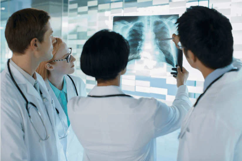
The use of Artificial Intelligence (IA) is transforming medical care in laboratories, clinics and hospitals. Through the use of technology, patient care can be improved, laboratory analysis processes can be optimized and diagnostic imagingas well as offering more efficient hospital management.
Artificial intelligence uses various algorithms that enable highly complex reasoning processes to be carried out, automating many tasks and functions. The use of AI in medicine provides multiple benefits and has a key role to play in the implementation of disease prevention and diagnosis, search for novel treatments and improvements in patient prognosis.
In the following article, we explain the process for implementing Artificial Intelligence solutions in laboratories, clinics and hospitals and the different applications that currently exist.
How to implement AI in laboratory and hospital analysis
Before starting to use artificial intelligence in the clinical setting, it is important to have an well-defined and structured strategy that integrates the technology along with the correct development of the process. These are the main steps to implement AI effectively:
1. Define the main objectives
The first step is to establish the objectives to be achieved with the integration of AI in the healthcare center. Among them, we can highlight:
- Reduction of diagnostic times.
- Customize treatments.
- Optimize resource management.
- Improve patient experience and care.
By setting clear goals, specific solutions can be provided using artificial intelligence, which will enable optimize healthcare management and save time and resources.
2. Analyze weaknesses and needs
Once the main objectives have been set, it is essential to carry out a complete diagnosis of the laboratory, clinic or hospital to analyze its weaknesses. This analysis should include the review of workflows the identification of the most important main problems and the areas that have a greater administrative or technical burden.
On the other hand, it is also important to involve medical, administrative and technical personnel in this process, as their day-to-day experiences provide a more accurate picture of real needs. Through a collaborative approach, AI solutions will be aligned with the specific challenges faced by the organization.
Select the right AI tools and solutions.
Subsequently, you must selecting the artificial intelligence technologies best suited to the hospital area. AI tools are revolutionizing the healthcare sector, especially in hospitals and laboratories, by improving diagnostic accuracy, increasing operational efficiency and delivering better healthcare. In this process, it is important to research the options available in the market and work with specialized healthcare technology providers.
Ensure proper integration into the healthcare ecosystem.
For the successful implementation of AI, it is crucial that new technologies are integrated with the systems that were already in use previously. Some of the tools we can highlight are hospital management software and its linkage with medical equipment, as well as the RIS system and the PACS system.
One of the essential aspects to achieve a correct integration is the interoperability concept. It refers to the importance of systems being compatible and capable of sharing information so that they can work in a coordinated and joint way in the different processes. For this reason, before applying the use of artificial intelligence, it is necessary to check that the systems to be used are compatible with each other.
5. Staff training
Another element to be taken into account is to provide a adequate staff training who will work with these technologies. This includes both medical and administrative staff, as they will be in charge of managing the tools, interpreting the data provided by AI and making the most of them in their day-to-day work.
In addition, it should fostering a culture of trust in technologyHe stressed that AI will not replace professionals, but is a tool that complements and improves their work. With this, it will be possible to ensure a correct transition to the application of new processes and innovations.
6. Ensuring data security and privacy
The management of medical data involves a great responsibility in terms of security and privacy. The implementation of AI must comply with local and international regulations, such as the General Data Protection Regulation (GDPR) in Europe. In this way, it will be possible to ensure that patient information is protected at all times.
The main measures include the correct data encryption, a user authentication and the anonymization of information whenever possible. In addition, it is crucial to conduct regular audits to identify and correct possible vulnerabilities in the systems.
7. Implement in a staggered manner
Introducing AI gradually is a fundamental strategy for minimize disruptions to day-to-day operations and facilitate staff adaptation. You can start with a pilot project in a specific unit, such as radiology, and evaluate its impact before extending the implementation to other areas.
During this phase, it is important to collect feedback from staff and adjust the tools according to their needs and suggestions. Through this step-by-step approach, improvements can be made progressively and achieve a adequate adoption of new artificial intelligence tools.
8. Monitor and measure results
The implementation of AI must be accompanied by an continuous monitoring to ensure that the solutions are meeting the established objectives. This involves define key performance indicators (KPIs)The results of this study have been significant, such as a reduction in diagnostic time, an increase in operational efficiency and an improvement in patient satisfaction. Regularly evaluate these results will identify areas for improvement and adjust strategies as needed, taking full advantage of the benefits of artificial intelligence in healthcare.
9. Promote continuous innovation
The implementation of AI is not an isolated action, but rather a continuous process. Technology is a sector that is constantly evolving. Therefore, it is important to be aware of new tools and methods in the healthcare area in order to be able to implement future improvements. To ensure that a medical institution is committed to innovation and is competitive in its sector, the following can be done to promote various actions. Among them, we can highlight:
- Foster a culture of innovation among the personnel.
- Participate in research programs.
- Collaborate with universities or technology companies.
- Implement new tools and methods.
Artificial intelligence solutions for laboratory analysis, clinics and hospitals

Source || Freepik
What kind of solutions can be implemented to optimize clinical and hospital management?
Software with artificial intelligence
Through the use of a AI softwareon the same platform, you can storing medical images generated in diagnostic imaging studies, manage patient data in real time, generate automated reports and make comparisons of current studies with previous medical imaging.
AI-assisted diagnostic imaging
The current medical equipment can integrate diagnostic imaging software with AI. These systems employ advanced algorithms to identify anomalies and diseases early, improve diagnostic accuracy and reduce analysis time. They can be used for different types of equipment, ranging from X-raysCT scans, CT scans or TAC, ultrasounds y mammograms to magnetic resonances.
Virtual agents for laboratory analysis and hospital centers
A virtual agent provides automation of different tasksIt can therefore be implemented in the healthcare sector to optimize the management of medical centers, clinics and laboratories. Through an artificial intelligence platform such as Serenity Star AIcan be implemented chatbots and virtual assistants that offer 24-hour patient support, improving customer service. Among its advantages, it stands out for providing instant information on hospital services, resolving patient queries, guiding patients in their search for specialists, and managing appointments and other administrative procedures.
The use of virtual agents also offers other functions very useful in research and hospital management. They allow the analysis of complex medical data with high accuracywhich allows accelerate the performance of medical studies and develop improvements and innovations in areas such as research and laboratory analysis.
Laboratory process automation
There are AI systems that allow automate many functions in laboratory analysis processes. From the performance and analysis of clinical tests to inventory management and the implementation of quality control improvements. Its use helps reduce human error, increase operational efficiency and reduce study processing time.
AI-assisted surgical robots
In the field of surgery, AI and robotic systems are making a difference. The use of AI-assisted surgical robotsas Da Vinci, help to realize more precise and less invasive procedures, decrease surgical risk and reduce recovery times of patients.
Another of the most noteworthy advances in this area is the creation of surgical simulation models to plan, practice and refine procedures before performing them in clinical practice.
Advances in telemedicine: Use of portable and AI-integrated medical equipment
Among the latest innovations, we can highlight the development of portable and AI-integrated medical devices. Its use offers continuous monitoring of patients outside the hospital environment, achieving great advances in telemedicine.
The telemedicine is one of the most outstanding areas of medical innovation, as it allows for assisting people with chronic diseases remotely and reach regions where medical services are not fully available. In this way, regardless of the specialist's location, fast and accurate diagnoses can be made.
Implementing artificial intelligence in laboratories, clinics and hospitals is a process that requires planning, collaboration and a strategic vision. From identifying needs to monitoring results, each step is crucial for ensure that AI is integrated effectively and generates tangible benefits. With proper execution, AI can transform healthcare, improving service quality, optimizing resources and ushering in a new era in healthcare management.
Contact with us to implement AI in the hospital environment.
Bibliography
Castro Beltrán, J., Vivas Gamboa, R. C., & Caicedo, J. (2023). Artificial intelligence in medicine: A narrative review on advances, applications and limitations.
Risaralda Medical Journal, 29(2), 101-110. Retrieved from
https://ojs2.utp.edu.co/index.php/revistamedica/article/view/25606
Díez-Peña, E. (2023). Artificial intelligence in medicine: present and future. Andalusian Journal of Medical Electronics and Robotics, 8(4), 30-37. Retrieved from https://www.rade.es/imageslib/PUBLICACIONES/ARTICULOS/V8N4%20-%2012%20-%20CON%20-%20DIEZ_IA%20medicina.pdf
Martínez-González, L. (2023). Applications and challenges of artificial intelligence in the medical sector. Journal of Medicine and Health, 15(3), 45-55. Retrieved from https://remus.unison.mx/index.php/remus_unison/article/view/178
Kiko Ramos
CEO of 4D Médica. Expert in marketing and distribution of medical equipment.

por Kiko Ramos | Jan 17, 2025 | Equipment analysis
The radioprotection is the set of measures, standards and practices aimed at protecting people, the environment and the surroundings from the harmful effects of ionizing radiation. In the clinical setting, the aim of radiation protection is to ensuring the safe use of radiation for diagnostic and therapeutic procedures for patients and healthcare personnel, minimizing the associated risks.
What is radioprotection?
The ionizing radiation is a fundamental tool in modern medicine. It is used in procedures of diagnostic imaging that use X-rays, such as conventional radiography, digital radiology, fluoroscopy, computed tomography (CT) and interventional radiologyRadiology, a branch of radiology that diagnoses and treats various pathologies by means of minimally invasive procedures. In turn, it is also used in radiotherapy treatmentsThe aim of this program is the destruction of tumor cells and tissues by means of radiation, and in the nuclear medicine.
However, its improper or excessive use can have harmful consequences for people's health.. These include tissue damage or increased risk of cancer in the long term. For this reason, it is of great importance in the clinical environment and requires a sound management. In this sense, the discipline of Radiation ProtectionThe company, which employs professionals such as physicists, physicians, biologists and engineers, is working to ensure that the development and application of technologies that use ionizing radiation are safe.
Basic principles of radiation protection
The Radiation Protection System is based on three fundamental principles that have been established by the International Commission on Radiological Protection (ICRP):
1. Justification
Any procedure involving the use of ionizing radiation must be medically indicated. This means that the benefits of the procedure must clearly outweigh the risks associated with radiological exposure.
2. Optimization (ALARA Principle)
Exposure should be kept "as low as reasonably achievable". This principle is referred to as ALARA and ensures that the lowest dose necessary to obtain clinical results is used.
3. Dose limitation
Strict dose limits must be established to protect both healthcare personnel and patients, preventing exposure from exceeding levels considered safe. This principle is oriented to the protection of persons exposed to radiation sources.
Application of the Radiological Protection System in the clinical environment.
In the clinical environment, the Radiation Protection System is implemented through a structured approach that includes the following aspects:
Design and maintenance of installations
The rooms of X-raysCT scans, CT scans or TAC and radiotherapy must be equipped with adequate shielding that minimizes radiation scattering. In turn, it is essential to carry out periodic inspections to guarantee the correct functioning of medical equipment and that they do not emit an unnecessary dose of radiation.
Equipment quality control
The following must be implemented preventive maintenance and calibration programs to ensure that the equipment operates efficiently within the established limits. Another key aspect is incorporate advanced technologies that allow automatic adjustment of radiation doses according to the patient's characteristics. To this end, digital radiology medical equipment will make it possible to optimize the amount of radiation, increasing safety in the healthcare environment for both medical staff and patients.
Staff training
One of the strategies to promote radioprotection in the clinical setting is to to train health professionals on the safe use of medical equipment that emit ionizing waves and which, in turn, have knowledge of the three principles of radioprotection. In this way, through appropriate training, it will be possible to promote the development of a safety culture to ensure the application of good practices in daily work in the health sector.
Radiation protection measures
Radiation protection in the clinical environment is essential to ensure the safety of patients and healthcare personnel from the risks associated with ionizing radiation. To this end, various strategies and tools designed to minimize unnecessary exposure are implemented, respecting the principles of justification, optimization and dose limitation.
Protection of healthcare personnel
Personnel working in areas where ionizing radiation is used must be adequately protected to avoid cumulative exposure that may pose a long-term risk. Key measures include:
- Personal Protective Equipment (PPE)Professionals should wear leaded aprons, thyroid protectors, leaded goggles and gloves that are specifically designed to reduce direct exposure to radiation.
- Dose monitoringIt is mandatory for health personnel to record the amount of radiation accumulated. This monitoring ensures that the dose does not exceed the limits established by the regulations in force.
- Staff turnoverTo minimize exposure time, personnel rotation is organized in tasks involving the handling of radiation-emitting equipment. In this way, the exposure load is evenly distributed.
Patient protection
Patients should also be protected from unnecessary radiation exposure, especially considering that they are often exposed in a timely manner but with high doses in some diagnostic or therapeutic procedures. The most relevant measures are:
- CollimationIt is essential to limit the area of the body that is exposed to radiation by using collimation systems that focus the radiation beam only on the area of interest. This reduces the amount of tissue irradiated and thus the associated risks.
- Optimized protocolsModern equipment makes it possible to adjust the exposure parameters (such as energy and radiation time) according to the specific characteristics of each patient. This makes it possible to deliver a minimum dose without compromising the quality of the radiation dose. medical images or treatment.
- Repetition controlTo avoid unnecessary repetitions of radiological studies, it is essential that the staff is well trained and that the equipment is functioning optimally. This ensures that the images obtained are of diagnostic quality on the first attempt.
Signaling and delimitation of areas
Facilities using ionizing radiation must have proper signage and access control to protect those not involved in the procedures. These measures include:
- SignageVisible signs should be posted indicating radiological risk areas and exposure levels, warning people of the need to wear appropriate protection or to avoid entry.
- Delimitation of areasIonizing radiation: Access to areas where ionizing radiation is used should be restricted. Its use should be limited to authorized personnel, thus avoiding accidental exposure of third parties or the general public.
In conclusion, radiation protection in the clinical setting is a shared responsibility that requires the collaboration of professionals, patients and regulatory bodies. Applying protection principles and measures not only ensures safety, but also improves the quality of medical care.
Kiko Ramos
CEO of 4D Médica. Expert in marketing and distribution of medical equipment.
por Kiko Ramos | Jan 9, 2025 | Projects
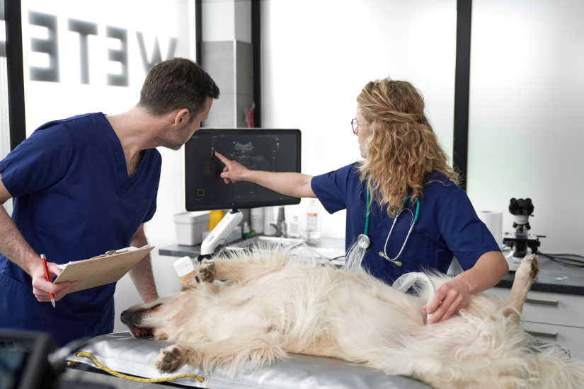
4D Médica has collaborated with the Hospital Clínico Veterinario CEU on the diagnostic imaging area. To this end, it has provided both the sale of different equipment like support, installation and maintenance of supplied medical equipment. The CEU Hospital is specialized in the area of Small Animals and Large Animals, so the cooperation with 4D Medical is a revolution in the clinical and research activity of the veterinary sector.
The CEU Clinical Veterinary Hospital, a reference center in the Valencian Community
The CEU Clinical Veterinary Hospitallocated in Valencia, has more than 20 years of experience in the veterinary field. This is in addition to the specialization of the medical team, new facilities, research and state-of-the-art technology. All these aspects have turned it into a reference center in the Valencian Community.
At June 2016was inaugurated on new Clinical Veterinary Hospital for Companion Animals and Large Animals at CEUof the Cardenal Herrera University. The center was built in a new facility with a total surface area of 4,536 square meterswhere they are housed diverse areas of expertise and spaces for clinical practice and research. Another aspect for which it stands out is that it is equipped with infectious animal isolation unitsbeing the first hospital in Spain that incorporates them.
The three pillars of the center
CEU Hospital focuses on three main pillars:
- TeachingAt the hospital, undergraduate and postgraduate students of the Faculty of Veterinary Medicine of the CEU Cardenal Herrera University carry out their internships, and continuous training is provided to health professors. The Faculty is a member of EAEVE (European Association of Establishments for Veterinary Education), an entity that brings together the best Veterinary Faculties in Europe.
- ClinicalThe center is a referral center for owners and veterinary professionals who refer the diagnosis and treatment of complex cases.
- ResearchResearch activities, both internal and in collaboration with external entities.
Multidisciplinary referral service
It offers a multidisciplinary referral service with qualified specialists with experience in national and international hospitals. The hospital is open 24 hours a day, 365 days a year to attend to veterinary emergencies, prioritizing a quality medical care.
Medical equipment and technology in the area of Diagnostic Imaging, provided by 4D Médica.
It is a center that incorporates the latest technology in Hospitalization and ICU Units. With the 4D Médica collaboration and the medical equipment provided in the area of Diagnostic Imaging, has become a pioneer and reference hospital in the veterinary sector. For this purpose, it has audiovisual and information systems that facilitate the transmission of operations from the operating rooms and a data processing center which manages information in real time, connected to the university network.
New Large Animals area
The new facilities are equipped with the clinical care services for large animals, especially equines. It is, therefore, in the largest equine hospital in the Valencian Community. The diagnostic services offered are digital radiology, ultrasonography and video endoscopyamong other techniques. Regarding the type of health care, within the area of large animals, the following are carried out emergency, general medicine, specialized internal medicine, surgery and anesthesia clinical services.
Medical equipment and services supplied by 4D Médica
In the cooperation between 4D Médica and the Hospital Clínico Veterinario CEU in the area of Diagnostic Imaging, the following medical equipment was supplied:
Digital X-ray detector Vivix V-2430VW (2018)
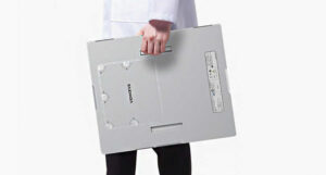
During 2018, 4D Médica provided the detector for. X-ray panel Vivix V-2430VW. It is a digital flat-panel detector of X-raysdesigned for general radiography applications. It is part of the Vieworks VIVIX-S V series, recognized for its advanced technology and robust design. Among its main features, we can highlight the following elements:
- Superior image qualityWith a pixel size of 124 µm, it offers a high spatial resolution of 4.0 line pairs per millimeter (lp/mm), capturing sharp, detailed images with minimal noise, enabling more reliable and accurate diagnoses.
- Ergonomic, lightweight and durable designThe detector is easy to maneuver and lightweight, making it comfortable to use for radiology technologists. In addition, it has built-in handles for easy carrying. At the same time, its glass-free design makes the detector resistant to shocks and drops, ensuring a long-lasting investment. In addition, it has an IP67 rating, which means it is dust and water resistant.
- Prolonged autonomyVIVIX-S 2430VW batteries last up to 16 hours on a single charge, allowing for intensive use without interruption. It supports a variety of charging methods, including wired charging, wireless charging and charging via a docking station.
- Automatic Exposure Detection (AED)Vieworks' patented Automatic Exposure Detection (AED) technology ensures that images are captured at the appropriate radiation dose for each patient, optimizing safety and comfort.
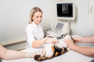
Sonosite M-Turbo, Sonosite Edge II, Wisonic Piloter and SIUI Ultrasound Scanners (2019-2024)
From 2019 to 2024, different models have been supplied and types of ultrasound scanners for accurate and quality diagnostic imaging.
Source || Freepik
Sonosite M-Turbo
It is a portable ultrasound scanner that is recognized for its durability and reliability. Weighs approximately 3.4 kg with batteryThis makes it easy to transport. It is designed for demanding environmentsIt offers a wide range of products and services, meeting U.S. military specifications. advanced image quality and turns on in less than 20 secondsIt can be used immediately. It is used in various diagnostic applications and clinical procedures around the world. From remote hospitals to veterinary clinics and community health centers.
Sonosite Edge II Ultrasound Scanner
This portable ultrasound system provides an enhanced imaging experience, while maintaining the same high quality of the images. design pillars: durability, reliability and user-friendliness. It has a wide-angle screen with an anti-reflective coating and has a simplified interface with a tactical panel with selection button that provides efficient navigation. At the same time, its silicone keyboard is sealed to prevent the entry of liquids and to allow adequate disinfection. On the other hand, it is ready to scan in less than 25 seconds and is suitable for various clinical applications.
Wisonic Piloter
It is a extremely lightweight tablet ultrasound scanner and with a weight of only 2 kg. It has a 13.3-inch touch screen that allows horizontal and vertical viewing. Through its extremely lightweight housing, intelligent design and built-in customized workflow, Piloter provides a more efficient experience for the user. musculoskeletal control, pain management, physical therapy and vascular system. Includes the wiNeedle technology for improved needle visualization during interventional procedures.
SIUI
The SIUI brand offers a variety of portable ultrasound scanners and with support for various clinical applications. For example, the Apogee 5300 Pro model is prominent in cardiology and radiology, offering advanced informatics capabilities that enable automated measurements essential for diagnosis. These devices are designed to provide a satisfactory image quality and intelligent applications that enhance diagnostic quality.
Portable X-ray generator MEX-20 (2022)
In 2022, the CEU Clinical Veterinary Hospital was awarded the MEX-20 portable X-ray generator. It is a portable high frequency X-ray generator, specifically designed for veterinary applications, especially in equine. It has the following components:
- Power and performancePower rating of 1.6 kW, equivalent to 4 kW in conventional systems, with a range of 50-90 kV adjustable in 1 kV increments, and a current of 20 mA.
- PortabilityNet weight of approximately 6.8 kg (including battery), it is easily transportable, facilitating its use in different clinical and field environments.
- AutonomyThe system is equipped with a high capacity battery (1600 mAs), which allows up to 160 exposures at 80 kV/10 mA on a single charge. Recharging time is less than 3 hours, ensuring continuous operation.
- Ease of useIt has an intuitive touch screen for parameter settings and has a memory for storing up to 10 presets. In addition, it has a dual laser pointer that facilitates precise positioning.
- Included accessoriesThe equipment includes a remote control and a portable case, providing protection and ease of transport.
Maintenance and repair of conventional small animal X-ray and endoscopy room (2017-2024).
Since 2017, in addition to the medical equipment provided, there have also been. maintenance and repair services for conventional X-ray room equipment of small animals and endoscopy.
Radiological image storage PACS, with 4D Medical installation and maintenance (2019-2024).
Source || Freepik
Technology is a key aspect that fosters better management of medical information and images. Starting in 2019, 4D Médica included the PACS system in the radiology area of the Hospital Clínico Veterinario CEU. This is a computerized image archiving and communication system which is used in the radiology area to store and manage reports and medical images electronically. With this, the hospital has a data processing center that manages data in real time and is connected to the university network.
Through this project, 4D Médica and the Hospital Clínico Veterinario CEU have driven the medical innovation and cutting-edge technology in the field of diagnostic imaging. This has allowed us to provide a quality clinical service, teaching and research, becoming a pioneering and widely recognized veterinary center in the Valencian Community.
Bibliography
CEU Cardenal Herrera University (n.d.). Presentation of the CEU Clinical Veterinary Hospital. Retrieved January 9, 2025, from
https://www.uchceu.es/hospital-clinico-veterinario/conocenos/presentacion
Sonosite (n.d.). M-Turbo: Portable ultrasound system. Retrieved January 9, 2025, from. https://www.sonosite.com/la/products/sonosite-m-turbo
Sonosite (n.d.). Edge II: Portable ultrasound system. Retrieved January 9, 2025, from. https://www.sonosite.com/es/products/sonosite-edge-ii
Wisonic (n.d.). Sample Page - Wisonic Spain. Retrieved January 9, 2025, from https://wisonic.es/index.php/sample-page/
SIUI (n.d.). SIUI products for diagnostic imaging. Retrieved January 9, 2025, from. https://www.siuivet.com/
Vieworks (n.d.). VIVIX-S 2430VW: Digital X-ray detector. Retrieved January 9, 2025, from. https://xrayimaging.vieworks.com/es/detector/radiography/1231
4D Medical. (n.d.). Mex20HF Plus portable RX Mex20HF Plus equipment. Retrieved January 9, 2025, from. https://www.4dmedica.com/equipo-rx-mex20hf-plus-portatil-p-1-50-1722/
Kiko Ramos
CEO of 4D Médica. Expert in marketing and distribution of medical equipment.

por Kiko Ramos | Jan 3, 2025 | News
Substrate AIa Valencian artificial intelligence company listed on BME Growth, has hired the consulting firm LKS Next to prepare the IPO of its subsidiary 4D Médica.
LKS Next is part of the Mondragon Corporation and has a total of 800 professionals specialized in the industrial, technological and health sectors. Its experience includes mergers, acquisitions and capital markets advisory servicesand, therefore will assist Substrate AI in preparing for the IPO.. From the preparation of the necessary documentation, the choice of the most suitable market for the initial public offering (IPO) and the search for investors, considering the particularities of 4D Médica's business.
Acquisition of 4D Medical by Substrate AI
Substrate AI acquired the 70% from 4D Medical in 2022 for 1.4 million euros. 4D Médica was originally dedicated to the sale of diagnostic imaging hardware for the veterinary sector.under the direction of its founder and CEO, Francesc Ramos, with more than 20 years of experience in the sector.
After the acquisition, the company was transformed into a AI applied to diagnostic imagingwith hardware and software divisions, operating in both veterinary and human medicine.
The purchase of Diagximag is integrated into the subsidiary 4D Médica
In 2023, Substrate AI purchased Diagximaga company focused on hardware for human medicine and main distributor of Samsung in Spain.integrating it with 4D Medical. In addition, we developed a AI-based imaging software to help doctors and veterinarians obtain more accurate diagnoses, which will soon also be available for human medicine.
This software seeks to Improve disease diagnosis and reduce radiation exposure to patients and physicians. by means of collimator self-regulation in ionizing radiation equipment.
4D Médica's sales growth since 2021
Thanks to these initiatives, 4D Médica has tripled its sales in three years1.8 million in 2021 to a figure three times higher in 2024, while maintaining an EBITDA margin of more than 25%.
"We are very satisfied with the road we have traveled in just two years. Together with Substrate AI, and thanks to their technology and support, we have transformed the company and prepared ourselves to become one of the key players in the application of AI to diagnostic imaging, an essential part of current and future medicine," says Francesc Javier Ramos, CEO of 4D Médica.
Therefore, the next step is the joint work of LKS Next, Substrate AI and 4D Médica to ensure that the IPO fits the needs of the company's growth plan.
Kiko Ramos
CEO of 4D Médica. Expert in marketing and distribution of medical equipment.
por Kiko Ramos | Dec 27, 2024 | Equipment analysis
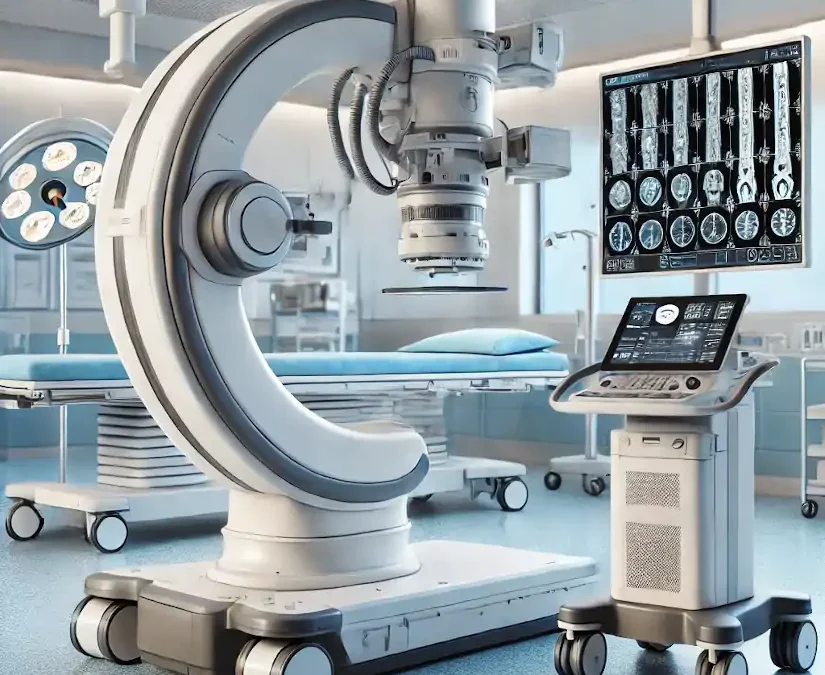
The arc in C is specialized medical equipment used in radiology and interventional procedures to obtain real-time X-ray images of the inside of the human body. It is a mobile device that enables radiological and fluoroscopic imaging. Its name derives from its C-shaped structure"which allows a wide range of movements and the acquisition of images from multiple angles and positions for capture specific anatomical views without moving the patient.
It is used to obtain X-ray and fluoroscopic images without having to move the patient to the radiology department. Therefore, diagnostics and procedures can be performed at the patient's hospital bedside or on the operating table during surgery. Its use is essential in areas such as surgery, orthopedics, traumatology, cardiology, neurology, urology and minimally invasive procedures.
Among the main advantages offered by the arc in Cis that facilitates diagnosisoffers a high precision and safety, y decreases the duration of surgical interventions in which the patient is under general anesthesia. In the following article, we analyze how a C-arm works, its parts, functions and main applications and uses. medical equipment.
How does a C-arc work?
The operation of a surgical C-arm is similar to that of the X-ray machines conventional. Combine two main elements that work in an integrated manner How does this process work?
X-ray generator
The process begins with the X-ray tubelocated at one end of the "C" arm. This component emits a beam of radiation which passes through the patient's body. Collimators, which are adjustable devices on the tube, delimit the radiation field, ensuring that only the area of interest is irradiated. This not only improves image quality, but also minimizes radiation exposure to other areas.
When the X-ray beam passes through the patient's body, interacts with the different tissuesgenerating a phenomenon called differential absorption. The Denser tissues, such as bones, absorb more radiation. and are represented as white areas in the image. On the other hand, the soft tissues and air-filled areas allow the rays to pass through more easily, appearing in gray or black tones. This difference in absorption is what creates the contrast in radiological images.
Image detector or intensifier
At the opposite end of the X-ray tube is the image detector or intensifier. This component receives the rays that have passed through the patient and converts them into electrical signals. Modern detectors, called digital flat panel detectors, process these signals to generate high-resolution images. This advance has largely replaced traditional intensifiers, offering greater sharpness and less radiation exposure.
The signals captured by the detector are sent to a processing system that converts the data into digital images.. This software automatically optimizes parameters such as contrast, brightness and sharpness to ensure that images are clear and easy to interpret. These images are displayed in real time on monitors connected to the system, allowing the medical team to observe the area of interest while the procedure is being performed.
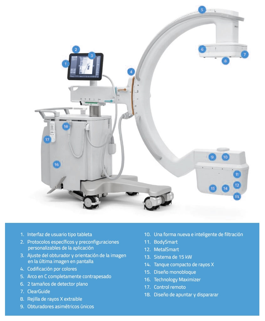
C-arc: Parts and functions
The C-arm in radiology consists of several parts that work together to provide high quality images in real time during medical procedures. Below are its main components and functions:
| Part |
Description |
| C-shaped arm |
Central structure connecting the X-ray tube to the detector. |
| X-ray tube |
Located at one end of the C-arm, it emits the radiation beam. |
| Image detector |
At the opposite end of the X-ray tube, it captures the radiation passing through the patient. |
| Mobile base |
Wheeled structure that supports the equipment and facilitates its transport. |
| Control panel |
Operational console from where the equipment parameters are adjusted. |
| Monitors |
Screens connected to the image processing system. |
| Collimator system |
Adjustable device located in the X-ray tube. |
| Cooling system |
Components that dissipate the heat generated by the X-ray tube. |
Parts of a C-arc
1. "C" shape arm
It is the main structure that connects the essential components of the equipment, such as the X-ray tube and the imaging detector.
Functions:
- The C-shaped arm connects the X-ray tube, which is located at one end, to the image detector or intensifier, which is located at the opposite end, allowing a wide range of movement around the patient.
- Facilitates imaging from multiple angles no need to move the patient.
- Includes rotations in multiple planes: horizontal, orbital and verticalThis makes it possible to adapt to different types of procedures.
X-ray tube
This is the radiation generator located at one end of the C-arm.
Functions:
- Emits X-rays through the patient's body.
- Its intensity and duration are controlled to obtain quality images. while minimizing radiation exposure.
- Security is a key aspect in the use of the C-arm. These devices are designed to minimize radiation exposure for both the patient and the medical staff. They have specific systems that reduce scattered radiation and integrated dosimeters continuously monitor the delivered dose.
3. Image intensifier or digital flat detector
It is located on the opposite side of the X-ray tube, capturing the radiation passing through the patient.
Functions:
- Converts X-rays into visible images in real time.
- The state-of-the-art digital flat panel detectors offer higher resolution images and lower radiation exposure compared to traditional intensifiers.
4. Control Console
This is the external control panel operated by the radiological technician during diagnosis.
Functions:
- Allows adjustment of exposure parametersThe company has a wide range of products, such as time and intensity, among other aspects.
- Controls the movement of the arc and the orientation of the images.
- Saves and transmits the images obtained for further analysis. The data is stored in a PACS system (Picture Archiving and Communication System), allowing quick and easy access for further analysis.
3. Monitor
The C-arm includes one or more high-resolution monitors, usually in Full HD, which allow physicians to view images in real time during procedures. This screen is connected to the system, usually located near the surgical field.
Functions:
- Displays radiological and fluoroscopic images in real time so that physicians can be guided through the procedure.
- Some systems include dual monitors to compare images in real time with previous analyses.
6. Mobility system
It is a rolling base with lockable wheels or fixed support system on larger models.
Functions:
- Facilitates C-arm transport between different areas of the hospital.
- Allows you to position the equipment in a stable and safe manner around the patient.
7. Power generator
It provides the power needed to operate the X-ray tube and other system components.
Functions:
- Regulates the power supply to ensure consistent performance during use.
8. Image processing software
By means of a radiodiagnostic softwareThe computerized system manages the acquisition, processing and storage of medical images.
Functions:
- Improved image quality through techniques such as contrast adjustment and noise reduction.
- Allows measurements and annotations directly on the images.
9. Collimator system
It is the device located in the X-ray tube that is responsible for controlling the irradiated area to be analyzed or treated.
Functions:
- Adjusts the radiation field to focus only on the area of interest.
- Reduces unnecessary radiation exposure for both the patient and the medical staff.
10. Refrigeration system
The cooling system is the mechanism for dissipating the heat generated by the X-ray tube.
Functions:
- Maintains equipment temperature within safe operating limits.
- Prolongs X-ray tube lifetime.
Clinical uses and applications of a C-arm in radiology
The C-arm is a medical device widely used in radiology and interventional medicine due to its ability to generate real-time images with high precision. What are its main uses and clinical applications?
Orthopedic surgery
In the field of orthopedic surgery, the C-arm is essential for the precise placement of screws, intramedullary nails and plates used in orthopedic surgery. fracture treatment. It is also used for guiding fracture reduction or deformity correction procedures. Its ability to provide clear, real-time images allows the surgeon to visualize bone structures and ensure that implants are positioned correctly, reducing the risk of errors during surgery.
Spine surgery
In spinal interventions, the C-arm facilitates the precise placement of the fixation devices such as pedicle screws and spinal fusion brackets. In turn, it is also used in procedures such as the vertebroplasty. The real-time images it generates are crucial to avoid injury to sensitive nerve structures and to ensure a successful outcome.
Interventional radiology
The C-arm is an essential tool in interventional radiology, where it is used for guided procedures such as biopsies, drains and tumor ablations. It is also indispensable in angiographieswhere digital subtraction imaging (DSA) allows high-precision visualization of blood vessels. This equipment facilitates minimally invasive procedures, which require detailed, real-time imaging to ensure accurate results.
Interventional cardiology
In cardiology, the C-arc is used in procedures such as coronary angiographieswhich evaluates the circulation in the arteries of the heart. It is also key to the implantation of pacemakers and other cardiac devices. Thanks to the dynamic images it provides, physicians can perform complex procedures with greater safety and precision.
Vascular surgery
In vascular surgery, the C-arm allows detailed visualization of the vascular system, which facilitates procedures such as the stenting to repair aneurysms or the insertion of filters in the vena cava.
Urology
In urology, this equipment is used to guide procedures such as placement of ureteral catheters or nephrostomies. It is also useful in the percutaneous nephrolithotomywhere kidney stones are removed using minimally invasive techniques. Real-time imaging helps physicians locate specific structures and avoid damage to surrounding tissues.
Gastroenterology
In gastroenterologic procedures, the C-arm is used for inserting feeding tubes or drainsas well as for placing esophageal prostheses. This device is especially useful in delicate procedures where precision is crucial, such as in hard-to-reach areas within the gastrointestinal tract.
Neurosurgery
In neurosurgery, the C-arm is used for procedures such as the electrode placement for deep brain stimulation or minimally invasive spinal surgeries. The ability to generate highly accurate intraoperative images is critical for navigating complex structures of the nervous system and ensuring patient safety.
Oncology
In the treatment of cancer, the C-arm is a valuable tool for radiofrequency or microwave ablationswhere localized tumors are destroyed. It is also used for the placement of markers to guide radiation therapy. Its ability to generate precise images allows for accurate positioning of instruments in malignant tissues, optimizing treatment.
Traumatology
In emergency situations or in traumatology, the C-arc is used for evaluate complex fractures and guide reduction procedures. It allows to verify in real time the correct alignment of the bones, which is crucial to ensure the patient's functional recovery.
Emergency procedures
In emergency environments, this equipment is indispensable for the immediate evaluation of serious injuriesas major trauma, and for guiding critical procedures such as thoracic drainage. Its ability to generate immediate images allows physicians to make quick decisions and save lives in critical situations.
Dentistry and maxillofacial surgery
In dentistry and maxillofacial surgery, the C-arm is used for the dental implant placement and surgical planning in the mandibular region. Provides detailed images of the bony structures of the skull and jaw, ensuring accurate results.
Gynecology and obstetrics
In gynecology, this equipment is used for interventional procedures, such as the placement of intrauterine devices or catheters used in fertility treatments. Its use improves the accuracy of procedures in sensitive areas, increasing safety and effectiveness.
Conclusion
The C-arm stands out for its versatility, as it is used in multiple medical specialties. Its ability to provide real-time imaging facilitates decision-making during complex procedures, reducing errors and improving clinical outcomes. In addition, by enabling minimally invasive interventions, it contributes to faster patient recovery and greater efficiency in medical resources.
If you are a health professional and you are interested in to acquire a C-arc or any other radiodiagnostic equipment, our 4D team will contact you to advise you and find the best solution for your clinic or hospital.
Contact 4D
Kiko Ramos
CEO of 4D Médica. Expert in marketing and distribution of medical equipment.
