por Kiko Ramos | Apr 4, 2025 | Equipment analysis
The fluoroscopy is a technique of diagnostic imaging which uses X-rays to observe the inside of the human body in real time. It is a type of X-ray that shows the internal structures of the organism in motion.. Unlike conventional X-rays, which generate static medical images, fluoroscopy creates dynamic images to analyze the functioning of different organs, tissues and other internal structures.
During a fluoroscopy, the fluoroscopea medical equipment that allows visualization of the patient's organs in motion. The dynamic images that are generated are projected on a monitor in video format so that medical professionals can diagnose and evaluate various medical conditions. This procedure is used to observe the structures and organs in operation. From seeing how the heart beats and how the lungs are inflamed to examining how food moves through the intestine. Therefore, it is very useful in studies of anatomy and physiology, as well as a support technique in certain interventions.
In the following article, we analyze fluoroscopy as a medical technique. From how a fluoroscopy examination is performed, the use of the fluoroscope and its different types to its main medical applications.
Fluoroscopy
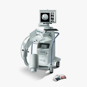
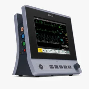
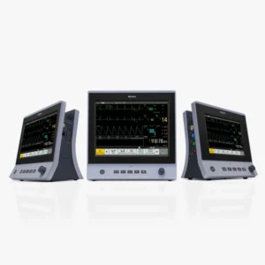
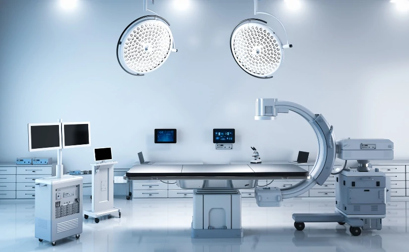
Fluoroscopy is a technique that allows you to see the inside of the body in motion and in real time. It combines X-ray technology, image detectors and digital processing to show what is happening inside the body. To do this, it is necessary to use specific medical equipment: the fluoroscope, also known as a C-arm.
Using continuous or pulsed X-rays, this device generates a set of dynamic images of the different organs, bones, tissues and joints in order to evaluate how certain structures of the body behave during a specific action. The different functions and parts of an arc in C allow radiological and fluoroscopic images to be taken.
Mainly, this medical equipment is used in a fluoroscopy examination for analyze the functioning of the organism when swallowing or breathing, as well as inspecting how a contrast liquid flows through the digestive or circulatory system. In turn, the fluoroscope is also used as a support technique in certain interventionssuch as stenting of blood vessels or cardiac catheterization.
How is a fluoroscopy procedure performed?
Although the visual result of fluoroscopy is a moving image, there is a certain process behind this technology. Understanding how it works is essential to evaluate its usefulness in medical diagnostics. Below, we explain step by step how a fluoroscopic examination is performed:
Patient preparation
In most cases, a very complex preparation is not necessary. Depending on the type of study, the patient will have to follow specific indications, such as fasting or temporarily suspending certain medications. Upon arrival at the medical center, the patient should taking off clothes, putting on a gown and removing metal objects such as necklaces, watches or belts, as they may interfere with the images.
Fluoroscopy examination
During the procedure, patient is positioned on a stretcher or standing in front of the fluoroscopethe team in charge of generating the dynamic images by means of the X-rays. The exploration consists of the following steps:
Administration of contrast medium
In many studies, a contrast medium is used to enhance the visibility of certain areas of the body. This contrast helps to highlight structures of interestallowing the physician to see with the functioning of the different organs and tissues more clearly.. This contrast can be administered in different ways, depending on the area to be studied:
- Oral routeIn case the area to be observed is the upper digestive system (esophagus, stomach).
- Intravenous lineWhen the examination is performed to evaluate the blood vessels or internal organs.
- Through a catheterFor bladder or bowel studies.
2. Capture and acquisition of images in real time
Once the contrast has been administered (if necessary), the technician or physician will begin capturing the medical images in real time. Throughout the procedure, it is important that the individual remains as still as possible.. Movement can distort the images, so the patient's cooperation is essential to obtain accurate results. During this phase, the specialist will be able to evaluate:
- The movement of an organThe diaphragm when breathing or the bladder when emptying.
- The passage of a contrast mediumto identify blockages, leaks or reflux in the digestive or urinary systems.
- The position of medical devicessuch as catheters, pacemakers, screws or prostheses.
- The dynamic function of a jointuseful in traumatology and physiotherapy.
This functional and dynamic approach is what distinguishes fluoroscopy from other imaging techniques, such as radiography or computed tomography (CT).
3. Medical image analysis
Modern fluoroscopy equipment is equipped with advanced technologies that enhance the analysis of medical images:
- Digital image processingDigital systems allow you to adjust different elements of the image, such as brightness, contrast, zoom and orientation.
- Recording and archivingThey offer the possibility to document the procedure or to review key sequences.
- On-screen measurementThe technology can be used to calculate lengths, angles or displacements automatically.
- Image overlay (fluoro overlay)It is very useful in image-guided interventions.
In addition, more and more systems are integrating artificial intelligence functions to assist in the automatic detection of anomalies or to improve visual quality in real time. Among the main advantages of using an AI software is that it increases diagnostic accuracy and facilitates medical decision making.
Duration of a fluoroscopic examination
The duration of the study may vary depending on the type of examination, the area to be explored and the complexity of the procedure. However, in general terms, a fluoroscopy usually lasts between 30 minutes and one hour. Once the examination is completed, the patient can return home and, in most cases, resume normal activities, unless otherwise instructed by the physician.
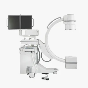
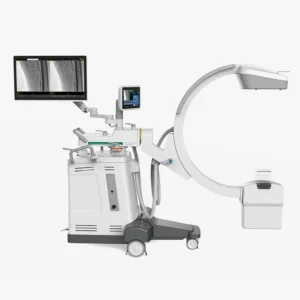
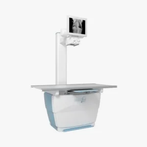
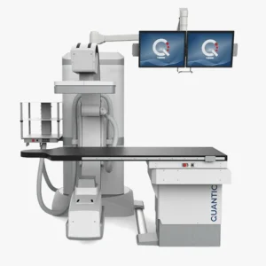
The fluoroscope: Types and characteristics
The fluoroscope, also referred to as a C-arm, is the medical equipment used in a fluoroscopic examination. However, there are different types of fluoroscopes depending on the type of study to be performed and the space available in the clinic or medical center. We can find two modalities and each one has specific characteristics:
| Features |
Full-Size C-Bow |
Mini C-Bow |
| Size |
Large, robust |
Compact, portable |
| Power |
High, for deep structures |
Medium/low, for surface structures |
| Main applications |
Orthopedic, vascular, spine, cardiac surgery |
Extremity surgery, hand, foot, pediatrics |
| Mobility |
Limited, requires more space |
Tall, easy to move and position |
| Complexity of use |
Advanced, requires technical training |
Simple, faster operation |
| Cost |
Higher |
More economical |
Full-Size C-arms
Full-Size C-Arches are designed to cover a wide range of procedures, from the simplest to the most complex.
- Large size: They have a wider field of view and are characterized by a greater ability to adjust to different positions and angles.
- Ample powerX-ray penetration: They provide deeper X-ray penetration, making them ideal for scanning complex structures such as the spine, thorax or pelvis.
- Advanced technologyMany models incorporate advanced technologies, such as 3D reconstruction, surgical navigation and high-resolution image processing.
- Medical applications: This type of arc is common in trauma, neurosurgery, vascular surgery and cardiac surgery operating rooms, where maximum precision and constant visual control are required throughout the procedure.
Mini bows
Mini C-arms are intended for more localized and less invasive procedures.
- Compact size: Their small size is ideal for small operating rooms, outpatient clinics or specialized practices, as they are much easier to transport and handle.
- Procedures of superficial areas of the bodyThese machines are optimized to work on more superficial areas of the body, such as hands, wrists, feet and ankles.
- Sharpness and lower powerAlthough their power is lower compared to full-size models, they offer clear and detailed images of the extremities. Therefore, it is recommended for minor surgeries or low complexity orthopedic interventions.
- Fast and easy operationFluoroscopes of this type are simpler to operate, as they have shorter start-up and positioning times. This improves efficiency in work environments where there is a high flow of patients.
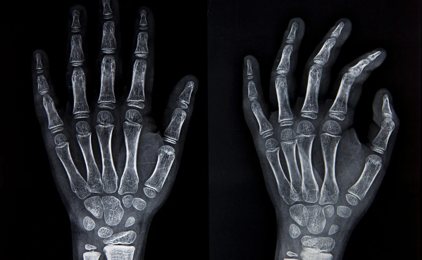
- Medical applicationsMini C-arms are especially useful in extremity surgery, outpatient trauma, hand and foot surgery, minor image-guided procedures and pediatric interventions.
Meet our 4D Medical equipment
What is fluoroscopy used for? Main medical applications
Fluoroscopy is used in many types of diagnostic imaging procedures. Among its main medical applications, we can highlight:

Examination of the digestive system
One of the most common applications of fluoroscopy is the study of the digestive system. By means of this procedure, the physician can observe how food or liquid moves through the digestive tract in real time. In this type of diagnostics, a contrast medium (such as barium) to be able to analyze more clearly the functioning of the esophagus, stomach or intestines.
Main applications
- Gastroesophageal reflux
- Hiatal hernias
- Ulcers or stenosis
- Swallowing disorders (dysphagia)
Cardiovascular system studies
In cardiology and interventional radiologyfluoroscopy is a key fundamental technique for visualizing blood flow through the heart and blood vessels. In these studies, fluoroscopy is used to act with greater precision during delicate interventions, reducing risks and complications. In addition, fluoroscopy is used to iodinated contrast agents that are injected intravenously to generate medical images of different tissues with greater clarity and sharpness.
Main applications
- Cardiac catheterization: Allows to see coronary arteries and detect obstructions.
- Angiography: Visualizes blood vessels in different parts of the body.
- Placement of stents or pacemakers: Fluoroscopy is used to guide the physician during these procedures.
Support in orthopedic surgeries and traumatology.
During bone or joint surgeryfluoroscopy helps surgeons to checking the position of pins, screws, prostheses or bone fragments. This allows interventions to be more precise and safe, reducing postoperative complications.
Main applications
- Spine surgeries
- Repair of complex fractures
- Image-guided joint infiltrations
- Arthrography (joint examination with contrast)
Minimally invasive procedures
Fluoroscopy is essential for performing image-guided procedures in which needles, catheters or probes are inserted into the body without the need for open surgery. By providing a real-time display, it allows to accurately access the area of interestThis reduces risks and improves the efficiency of the procedure.
Main applications
- Directed biopsies
- Abscess drainage
- Placement of central catheters
- Pain treatments (nerve blocks)
Use in pediatrics
Fluoroscopy, when performed in children, is used with reduced doses and special protocols to ensure its safety. Therefore, in the field of pediatrics, it is of great use for observe developing bodily functions.
Main applications
- Swallowing or reflux problems in infants
- Urinary tract malformations
- Evaluation of intestinal transit
- Follow-up of pediatric orthopedic surgeries
Functional evaluation of organs
In addition to detecting structures, fluoroscopy allows for the following see and analyze how certain organs function. In these cases, not only to detect abnormalities, but also to study how the body works in action.
Main applications
- To analyze how the bladder contracts during urination (cystography).
- Examine how the diaphragm moves when breathing.
- Perform an evaluation of gastric emptying.
Conclusion
The fluoroscopy procedure is a safe, non-invasive and highly effective technique for observing the body in motion. By combining X-rays and contrast media, medical professionals can obtain clear and accurate images that facilitate diagnosis and clinical decision making.
If you are looking for a fluoroscope for your clinic or hospital and you need more information, we help you to choose the medical equipment according to your needs. Contact us and we will answer all your questions.
Contact 4D Médica
Kiko Ramos
CEO of 4D Médica. Expert in marketing and distribution of medical equipment.


por Kiko Ramos | Mar 25, 2025 | Projects
4D Medica has collaborated with the Burgos and Valladolid Wild Animal Recovery Centersproviding medical equipment in the area of image diagnosis in the field of veterinary radiology. At present, in Castilla y León, there are three Wild Animal Recovery Centers (CRAS) in the provinces of Valladolid, Burgos and Segovia and two Wild Animal Reception Centers (CRF) in the provinces of Zamora and Salamanca. All of them are part of the network of wildlife care centers of the Natural Heritage Foundation of Castilla y León.
The CRAS have the primary function is to care for and recover injured wildlife species. with the aim of rehabilitating them and reintroducing them into their natural habitat. According to the Regional Ministry of the Environment, the last year has closed with record figures in the CRAS. Throughout 2024, a total of 8,600 specimens have been cared for in the network, an increase of 22%.
Among the different CRAS of the Community, Burgos and Valladolid are the provinces with the highest percentages of specimens received. According to data from the Junta de Castilla y León, Burgos has a total of 1,402 animals attended and Valladolid has 1,376 animals, representing 28% and 21% of the entries respectively.
The collaboration between 4D Medica and the animal recovery centers of Burgos and Valladolid arises through a public bidding by the Junta de Castilla y León (Castilla y León Regional Government). In this project, we have supplied different X-ray equipmentas well as protective equipment for professionals in the veterinary field.

The Burgos and Valladolid Wild Animal Recovery Centers
The Wild Animal Recuperation Centers in Burgos and Valladolid are managed by the Junta de Castilla y León and play a fundamental role in the wildlife protection, conservation and recovery. They are responsible for the care and rehabilitation of injured, sick or vulnerable wildlife, to later reintroduce them into their natural habitat.
The CRAS de Burgos was inaugurated on March 25, 2015. and is located in the town of Albillos, Burgos. Its facilities have more than 16,300 m² within a 47,000 m² plot of land and allow it to take care of a total of 100 birds and 25 mammals simultaneously. It is the largest recovery center in the Community of Castilla y León, making it a reference point for the provinces of Burgos, Palencia and Soria. For its part, the Wild Animal Recovery Center of Valladolid has been working with wildlife since 1989..
What are the main functions of recovery centers?
The CRAS of Burgos and Valladolid play an essential role in the specialized care of wildlife. In turn, they collaborate with institutions and scientific research projects on environmental and biodiversity conservation in the region of Castilla y León. Specifically, they perform the following functions:

Reception, veterinary care and rehabilitation of wild animals
Both centers receive wild animals from citizens, environmental agents or law enforcement agencies, where they undergo a thorough examination of the animals. veterinary diagnosis to identify injuries, wounds or diseases. Once at the center, the animal recovery in order to rehabilitate them and reintroduce them into their natural habitat in the shortest possible time, whenever feasible.
Breeding in captivity of protected or endangered species
They also participate in captive species breeding programs to reinforce wild populations or endangered animal species.
Biodiversity conservation
Contribute to the maintenance and recovery of native species and disease control which can affect wildlife, livestock and the human population, known as zoonoses.
Scientific research and health surveillance
Another of its functions is to collect data on the animals treated and deceased. From the different causes of admission, injuries and pathologies to the study of the evolution of the disease. In the Valladolid and Burgos centers, the following are carried out anatomopathological analysis of deceased specimensThis allows the detection of diseases, intoxications and other crimes against wildlife. These analyses also contribute to genetic censuses of protected species such as the Iberian wolf and the brown bear. At the same time, these centers participate in wildlife health surveillance studies and programs.
Environmental education and awareness
Through outreach activities, school visits or awareness-raising campaignspromote respect, respect for the environmental education and wildlife protection. Among their tasks, they are in charge of informing about the most common causes of entry of wild animals, such as roadkill, electrocution, illegal traps, etc.
Custody of species that cannot be reintroduced into the wild
Some animals cannot return to the wild by the presence of irreversible lesions. Thus, CRAS maintain and care for these animals. in the center and study the evolution of each one of them. for educational and scientific purposes.
Coordination with other entities
Recovery centers collaborate with the Territorial Environmental Service, police forces, research centers and other regional or state administrations. to protect wildlife and rehabilitate animals in need of veterinary care.
Medical equipment supplied by 4D Medica
In collaboration with the CRAS of Burgos and Valladolid, the following have been provided medical devices:
Two Digiray FireCR Spark X-Ray image acquisition systems
The FireCR Spark is a digital radiographic reading system developed by Digiray. This equipment is designed to provide high quality medical images in an efficient and adaptable way to various clinical needs.
Main features
- High image qualityThe FireCR Spark uses advanced signal collection technology that ensures clear and detailed images, facilitating accurate diagnoses.
- Compact and versatile designThe small size and light weight of the device allows it to be placed on a table or on the wall, optimizing space in clinical environments.
- Support for multiple cassette sizesThe system supports a variety of cassette sizes to suit different imaging needs.
- Adaptive processing speedThe FireCR Spark line offers models with different imaging speeds to meet the specific requirements of veterinary medical diagnostics.
- Advanced softwareQuantorMed+ Imaging software is included, providing an intuitive and easy-to-use interface. It offers fast and efficient operation and unlimited software updates, allowing the system to be kept up to date with the latest enhancements and functionalities.
Meet our medical team
Neovet X-Ray Equipment
Neovet equipment is a veterinary radiology system developed by the manufacturer Sedecal. It is specifically designed to meet the imaging needs of veterinary clinics and hospitals. This medical equipment combines advanced medical technology with functionalities adapted to the veterinary environment, facilitating high quality imaging for a variety of animal species.
Main features
- Versatility in different imaging modalitiesThe system offers both analog and digital solutions, adapting to the preferences and requirements of each clinic. In addition, it offers a choice of fixed or variable focal length and the possibility of angular projections, which expands the diagnostic options available.
- Exclusive STII Software (Only 3 Simple Touches)This intuitive software enables optimal digital imaging with as few as three interactions, streamlining the image acquisition process and improving efficiency in the clinical workflow.
- Control console optionsThe system offers three control console configurations to suit different spaces and preferences:
- Embedded tube support
- Pedestal
- Fixed to the wall
- Analog to digital upgrade kitFor clinics that initially opt for a conventional system, Sedecal offers an upgrade kit that converts the Neovet equipment to a digital system, allowing an easy transition to digital radiology and ensuring a good investment for the future.
Meet our medical team
Biochemistry equipment
A biochemistry team also called a biochemistry analyzer, is an automated device that can be used to analyze measures concentrations of chemicals in biological samples. Mainly blood and urine. These analyses provide information on the functioning of various organs and systems of the animalssuch as the liver, kidneys, pancreas or general metabolism.
Veterinary equipment is adapted to work with species-specific reference values, which is key in animals with very different physiologies. From dogs, cats, and horses to cattle or poultry.
Main functions in veterinary medicine
- Evaluate the liver function.
- Analyze the kidney function.
- Detect metabolic disorders.
- Electrolyte profile studywhere sodium, potassium, calcium and chlorine values are measured.
- Follow-up of treatments or surgeries
- Preventive control and routine check-ups
Hematology team
A hematology team is an automated analyzer that studies the cellular composition of animal blood.. By means of this device, it is possible to perform a complete hemogramadapted to the hematological particularities of different animal species. These devices use technologies such as electrical impedance, flow cytometry or colorimetry to count, classify and measure the characteristics of red blood cells, white blood cells and platelets.
Main functions in veterinary medicine
- Diagnosis of hematological diseases
- Follow-up of chronic or infectious diseases
- Pre-surgical evaluation
- Geriatric check-ups and preventive check-ups
- Rapid evaluations in field environments
Leaded X-Ray protection equipment for veterinarians
The X-ray shielding equipment with plumbed for veterinarians have the function of protect veterinary personnel and auxiliaries from exposure to ionizing radiation. during the performance of radiographic studies on animals. This protective equipment consists of a set of garments and elements made of lead-containing materials, which are designed for block or attenuate X-raysthus protecting the most sensitive organs of the human body.
Although the doses used in veterinary radiology are usually low and localized, cumulative exposure can represent a significant risk if appropriate radioprotection in the clinical environment.
Meet our X-Ray protection accessories
Conclusion
With this medical equipment provided by 4D Medica in the veterinary area, the CRAS of Burgos and Valladolid can improve diagnostic accuracy in the treatment, rehabilitation and reintegration of wild animals treated in both centers.
Kiko Ramos
CEO of 4D Medica. Expert in marketing and distribution of medical equipment.
por Kiko Ramos | Mar 12, 2025 | Equipment analysis
The interventional radiology (IR) is a specialized branch of the radiology area that combines techniques of diagnostic imaging with minimally invasive therapeutic procedures to diagnose and treat different diseases. In contrast to traditional surgical procedures, which require large incisions and long recovery times, interventional radiology allows for treat diseases without the need for open surgery. In this way, it stands out as an innovative discipline that reduces risks, recovery time and postoperative complications.
In the last decades, interventional radiology has experienced a great growth with the development of new technological advances both in imaging techniques and in medical equipment for the diagnosis and treatment of patients. interventional radiology. In the following article, we analyze what it consists of, its different types, as well as its main advantages and disadvantages.
What is interventional radiology?
Interventional radiology uses a series of diagnostic imaging technologies to guide therapeutic procedures with a high precision. The main modalities used are the following X-raysthe ultrasoundsthe computed tomography (CT) scans and the magnetic resonance imaging (MRI).
These techniques offer detailed information on the patient's anatomy and physiology in real timeThis allows medical professionals to visualize specific areas of anatomical structures and access them by making small incisions. To do so, they use specialized instruments such as catheters and needles. The use of high quality images and live viewing capability during procedures not only facilitates device placement, but also plays a key role in minimizing the risks associated with the procedure and reducing damage to healthy tissues.
IR is a medical discipline that is used for the treatment of treatment of various medical specialtiesThe company's main areas of expertise include oncology, cardiology, neurology, vascular radiology and musculoskeletal medicine. In turn, it has the capacity to offer less invasive procedures for patients who have certain risks in conventional surgery, such as the elderly, as well as patients with advanced pathologies or with a high surgical risk.
Interventional radiology procedures are performed under the following conditions local anesthesia, patients are awake during the procedure. Therefore, the risks that may arise from the application of general anesthesia are reduced. Another aspect to highlight is that most procedures are performed on an outpatient basis. In this way, patients can return home on the same day of surgery, reducing hospital costs and increasing the efficiency of the healthcare system.
The multiple advances in technology offer a great projection in interventional radiology. The integration of artificial intelligence in medicine and robotics has a special relevance in this discipline, which will increase precision and efficiency in the treatment of many diseases.
Types of interventional radiology
Medical technology continues to advance and interventional radiology plays a fundamental role in modern medicine. It is now used in different medical specialties and covers a wide range of therapeutic procedures. The main types of interventional radiology include vascular, oncology, musculoskeletal, gastrointestinal, urological, thoracic and gynecological, which we discuss below:

Guided image diagnosis
One of the main functions of interventional radiology is the diagnosis of diseases through image-guided procedures. In many cases, it is necessary to take tissue samples or drain fluids accumulated in the body to obtain an accurate diagnosis. Through the use of imaging techniques, these procedures can be carried out with a millimetric precision and without the need for invasive surgery.
Main diagnostic procedures
- Image-guided biopsiesBiopsies: Fine needles are used to remove tissue samples from organs such as the liver, lungs, thyroid or prostate. These biopsies make it possible to detect diseases such as cancer in its early stages.
- Percutaneous drainageWhen there is fluid accumulation due to infection or inflammation, catheters are placed to remove it without the need for major surgery.
- Puncture and aspiration of cysts or massesUsing a needle guided by ultrasound or tomography, physicians can remove cysts or reduce pressure in areas with fluid accumulation.
2. Vascular and endovascular treatment
The diseases of the circulatory systemsuch as arteriosclerosis, aneurysms and varicose veins can be effectively treated with interventional radiology techniques. In these cases, physicians use catheters and guidewires for accessing blood vessels and perform procedures that improve circulation or prevent serious complications. These treatments offer a less invasive alternative to conventional surgery, reducing hospitalization times and improving patients' quality of life.
Main treatments
- Angioplasty and stentingIn patients with blocked arteries, a balloon is introduced through a catheter to widen the blood vessel. Subsequently, a stent, a small metal device that keeps the artery open and prevents future blockages, is placed.
- Aneurysm embolizationWhen aneurysms, dangerous dilatations of the arteries, arise, micro-spirals or embolizing materials can be introduced to reduce the risk of rupture.
- Treatment of varicose veins and vascular malformationsSclerotherapy techniques are used to close abnormal veins and improve circulation, eliminating aesthetic discomfort and associated circulatory problems.
Clinical applications
- Peripheral arterial disease.
- Cerebral and arterial aneurysms.
- Stroke, cerebrovascular accident.
- Varicose veins and venous malformations.
3. Interventional Oncology
In the field of oncology, interventional radiology has opened up new possibilities for the treatment of cancer. canceras it allows the localized destruction of tumorsThe impact on healthy tissues and its side effects are reduced. Therefore, interventional oncology represents an important effective and less aggressive alternative to surgery.
Procedures in interventional oncology
- Percutaneous tumor ablationRadiofrequency, microwave or cryotherapy techniques are used to destroy tumors in the liver, kidney, lung and other organs without the need for open surgery.
- Chemoembolization and radioembolizationChemotherapeutic drugs or radioactive particles are administered directly into the blood vessels that feed the tumor, reducing its size and preventing its growth.
- Placement of catheters and central venous accesses.In patients requiring prolonged chemotherapy treatments, venous ports are inserted to administer the drugs more comfortably and safely.
Clinical applications
- Liver, lung and kidney cancer.
- Bone and soft tissue tumors.
- Palliative treatment in oncology.
4. Traumatology and pain management
Interventional radiology procedures are also essential for the management of chronic pain and the treatment of musculoskeletal injuries. These procedures improve the quality of life for patients by reduce pain and restore joint function without the need to resort to open surgery.
Most common interventions
- Joint infiltrations and nerve blocksAnesthetic and anti-inflammatory drugs are injected into joints such as the knee, hip or spine to relieve pain caused by arthritis or other conditions.
- Cementoplasty (vertebroplasty and kyphoplasty)In this type of procedure, a bone cement is injected into fractured or osteoporosis-damaged vertebrae to reduce pain and improve spinal stability.
- Aspiration of calcifications and drainage of articular cystsCalcium deposits in tendons or fluid accumulated in joints are eliminated, improving the patient's mobility and reducing pain.
Clinical applications:
- Osteoporosis with vertebral fractures.
- Herniated discs and chronic low back pain.
- Rheumatoid arthritis and osteoarthritis.
5. Gastroenterology and urology
IR can be used to treat diseases of the digestive and urinary tract.
- Placement of esophageal and biliary prosthesisStents are inserted in the esophagus, bile ducts or intestines to allow the passage of food or liquids in cases of obstructions caused by tumors.
- Percutaneous nephrostomyDrainage: A drainage tube is inserted into the kidney to decompress urinary obstruction in patients with kidney stones or tumors.
- Treatment of gastrointestinal bleedingEmbolization is used to stop bleeding from gastric ulcers or esophageal varices, avoiding emergency surgery.
Clinical applications in gastroenterology
- Esophageal, liver and pancreatic cancer.
- Hepatic cirrhosis with portal hypertension.
- Biliary obstructions and intestinal strictures.
Clinical applications in urology
- Urinary obstruction due to tumors or kidney stones.
- Varicocele and fertility problems.
- Benign prostatic hyperplasia.
6. Pulmonary and thoracic interventional radiology.
This specialty allows the diagnosis and treatment of thoracic diseases without the need for invasive surgical procedures.
Main procedures
- CT-guided lung biopsyLung tissue sampling for cancer diagnosis.
- Pleural drainage and pleurodesisElimination of fluid in the pleural space in cases of pleural effusion.
- Embolization of pulmonary arteriovenous malformationsClosure of abnormal blood vessels in the lungs.
Clinical applications
- Lung cancer and pleural diseases.
- Recurrent pneumothorax.
- Pulmonary vascular malformations.
7. Gynecology and obstetrics
In this medical specialty, the following treatments can be performed gynecological pathologies and pregnancy complications with image-guided procedures.
Main procedures
- Uterine fibroid embolizationNon-surgical procedure that reduces the size of fibroids without removing the uterus.
- Treatment of postpartum hemorrhageUterine arteries are occluded to stop severe bleeding after delivery.
- Pelvic abscess drainageGynecological infections elimination with percutaneous catheters.
Clinical applications
- Uterine fibroids and abnormal bleeding.
- Severe postpartum hemorrhage.
- Pelvic abscesses due to infections.
Meet our 4D Medical equipment
Advantages of interventional radiology
Interventional radiology offers many advantages and has transformed the treatment of many diseases, offering safer, less invasive procedures with shorter recovery times.
Minimally invasive procedures: Less risk and greater precision
One of the major advantages of interventional radiology is that it allows treatments to be performed without the need for open surgery. Instead of making large incisions, it uses small punctures in the skin through which catheters, microneedles and specialized devices are introduced.
This results in less damage to surrounding tissuesthere is a reduced risk of postoperative infections and you get a reduction of bleeding and scar formationimproving the patient's recovery.
Shorter hospital stay and faster recovery time
Interventional radiology procedures, being less aggressive to the body, allow the patient to recover in less time compared to conventional surgery. Many of the procedures are ambulatoryThe patient returns home after the procedure and is then hospital stay is reduced.
Another aspect to highlight is that the simpler and less invasive interventions. In this way, the decreases the consumption of painkillers since postoperative pain is less. At the same time, the patient can return to his or her daily activities and work in a shorter time, since the recovery times are shorter.
Less need for general anesthesia
Unlike traditional surgeries, which usually require general anesthesia, interventional radiology procedures are performed with local anesthesia and light sedation. This minimizes anesthetic risksespecially in patients with chronic diseases or advanced age. In addition to reducing the risk of complications, interventional radiology offers safer procedures for patients with cardiac or respiratory problems.
Highly accurate and efficient diagnosis and treatment
Interventional radiology uses real-time imaging to guide the placement of needles, catheters and other medical devices with extreme precision. The use of techniques such as fluoroscopy, ultrasound, computed tomography or magnetic resonance imaging offers different benefits:
- Helps reduce the margin of error in complex procedures.
- Increased success rate of oncology and vascular treatments.
- Reduces collateral damage in adjacent structures.
Alternative treatment for patients who are not surgical candidates
For many patients with advanced disease or high surgical risks, interventional radiology is the only viable treatment option. It is a alternative for people suffering from advanced diseaseshave severe comorbidities or for those who reject invasive surgical procedures.
Covers multiple medical specialties
Interventional radiology is not limited to a single medical specialty, but is also encompasses several areas. Therefore, it provides a versatile treatment for treating diseases in different organs and systems, since it its approach is multidisciplinary. In addition, it is a discipline that is constantly evolving, thus allowing the application of new applications and technological improvements.
Lower cost compared to traditional surgeries
Although some interventional radiology procedures may involve more expensive medical equipment, their total cost is lower than that of conventional surgery. Among the main factors that reduce its costs, we can highlight the following aspects:
- Reduced consumption of medical resources and hospitalization time.
- Medication is reduced administered to patients.
- Faster recovery.
Disadvantages of interventional radiology
Despite its many benefits, interventional radiology is not without its limitations and challenges. Although it represents a less invasive alternative to traditional surgery, there are factors that may limit its application or affect patient safety.
Limited availability and restricted access
One of the main challenges in interventional radiology is that not all hospitals and clinics have the necessary technology and trained specialists. to perform these procedures.
In rural areas or countries with fewer resources, patients may not have access to advanced imaging equipment or interventional radiologists, limiting the possibility of receiving these treatments. In these cases, patients will have to travel long distances to receive care, so many of them will have to opt for more invasive surgeries due to the unavailability of interventional radiology.
Another disadvantage is that not all health systems finance these procedures. and this may generate economic barriers that hinder access to this medical discipline.
Not all procedures are equally effective in the treatment of the disease.
Although interventional radiology offers effective solutions for many diseases, some procedures only control symptoms or slow disease progression, but do not completely eliminate the disease. Therefore, IR emerges as a temporary solution until the patient is able to undergo definitive treatment. On other occasions, some treatments must be repeated several times to increase its effectiveness.
Use of ionizing radiation in some procedures
Many interventional radiology procedures, especially those that utilize X-ray machines and fluoroscopy, expose the patient to ionizing radiation. Although the doses are usually low, repeated exposure may increase the risk to the patient.
What impact can it have on the patient? On the one hand, cumulative exposure over the years. could increase the risk of adverse effectsespecially in repeated procedures. In turn, in young patients or pregnant women, the risk-benefit ratio should be evaluated with caution.
Possible adverse effects and complications
Although interventional radiology is generally safer than surgery, it is not without risks and complications. As these are minimally invasive procedures, there is a possibility of adverse effects in certain patients:
- Hemorrhage at the puncture siteMay occur in procedures that require the insertion of catheters in arteries or veins.
- Allergic reactions to contrast mediumIn studies such as angiography and cholangiography, some patients may present severe allergic reactions to iodinated contrast material.
- Infection at the puncture siteAlthough less frequent than in conventional surgeries, there is still a risk of infection.
- Device migrationIn rare cases, a stent or embolization coil may dislodge and cause unwanted obstructions.
Recent discipline and limited availability of professionals
The success of interventional radiology depends largely on the skill and experience of the interventional radiologist. In contrast to traditional surgery, where surgeons have extensive experience, IR is a relatively new specialtyand, therefore, the availability of highly trained professionals is still limited.
Conclusion
Interventional radiology is a recent medical discipline that offers high precision, reducing the application of invasive treatments and open surgery. In recent years, it has had a great impact on modern medicine, improving the quality of life of patients and reducing postoperative complications, hospitalization times and health care costs.
Do you need more information about medical equipment? Contact us and from the 4D Médica team we will help you find the equipment that best suits the different needs of your clinic or medical center.
Contact 4D
Bibliography
Dalda Navarro, J. Á., Navarro Martín, M. T., Negre Ferrer, E., Negre Ferrer, C., Navarro Martín, A. B., & Dalda Navarro, V. (2024).
Interventional radiology: minimally invasive treatments under image guidance. Health Research Journal, 5(6). Retrieved from
https://dialnet.unirioja.es/servlet/articulo?codigo=9693488?
Lonjedo, E. (2019). Interventional radiology: as far as the image takes you. Annals (Reial Acadèmia de Medicina de la Comunitat Valenciana), (20). Retrieved from https://dialnet.unirioja.es/servlet/articulo?codigo=7710219
Kiko Ramos
CEO of 4D Médica. Expert in marketing and distribution of medical equipment.
por Kiko Ramos | Feb 25, 2025 | News
The European Society for Medical Oncology (ESMO) is an international organization that multidisciplinary professional organization which promotes research, education, and international collaboration in the cancer treatment in Europe and worldwide. Founded in 1975, it brings together physicians, researchers and health professionals who are responsible for implementing innovative strategies and developing medical advances in the area of oncology.
What are the latest advances that have been made in oncology in the last year? In the following article, we analyze the importance of ESMO in medicine and the most outstanding research in cancer treatment.
ESMO's role in medicine
ESMO plays a key role in cancer research worldwide. Among its main functions, it supports research into new therapies, the development of personalized medicine and the use of artificial intelligence in the detection of cancer and in the area of diagnostic imaging. It is responsible for the creation of various clinical guidelines to promote the medical education and research into innovative cancer treatments.
To this end, it organizes congresses, courses and scientific publications to update professionals and specialists on the latest trends in oncological treatments. At the same time, it also elaborates international guidelines and protocols for cancer diagnosis and treatmentThe company's work in this field has led to many advances: the following are some of the most important advances in the field In particular, its work in this field has led to many advances:
- Create and implement safer and more effective treatments.
- Promoting prevention by means of a early diagnosis of cancer.
- Promote a equitable access to cancer care.
- Improving quality of life of millions of patients.
4 new advances in oncology presented by ESMO
On an annual basis, ESMO organizes one of the most important oncology congresses in the world: the Congress of the European Society for Medical Oncology ESMO. It brings together researchers, professionals and world leaders in the field of oncology to present the latest discoveries in oncology therapies, prevention strategies and technological innovations in medicine.
The last event took place in Barcelona, Spain, from September 13 to 17, 2024, where it was possible to analyze the latest advances in cancer treatment. The following is a summary of what was new:
New studies in immunotherapy
As recently as 15 years ago, the prognosis for a patient with metastatic melanoma was very limited. There was no way to slow the progression of the skin cancer and their life expectancy was less than six months. However, at the beginning of the last decade, great advances were achieved with the development of immunotherapy.
What does immunotherapy consist of? It is a technique based on the stimulation of the body's own defenses to eliminate the malignant cells present in the body. Today, immunotherapy studies have achieved a survival rate of 10 years for a person with the same disease. Its favorable results have allowed it to be expanded to other tumors and, at present, it is also being used to treat other tumors. is used in some types of lung, bladder and breast cancer.
A decade later, it has become a fundamental therapeutic approach that is still under continuous development and research.. During the ESMO congress, a study was presented showing the long-term impact of immunotherapy. The publication showed that half of the patients with metastatic melanoma who had been treated with immunotherapy survived cancer-free for up to 10 years.
Another research presented at the congress was that this type of drugs raises the survival of the most aggressive breast cancer: triple negative.
2. Intelligent precision chemotherapy
One of the major innovations addressed at the ESMO 2024 congress is the ADC drug developmentwhich combine a monoclonal antibody with cytotoxic agents. These drugs allow targeting chemotherapy directly to tumor cellsThis increases efficacy and reduces side effects.
Currently, the intelligent chemotherapy, with greater precisionis one of the most outstanding advances in oncology treatment and cancer cure. The use of ADC drugs represents one of the most promising solutions for the treatment of cancer. to treat different types of tumors, applying lower doses of chemotherapy and with lower toxicity.
3. Artificial Intelligence applied to Oncology
Artificial intelligence (AI) is revolutionizing oncologyThe results of this research range from predicting responses to treatments to detecting genetic alterations that are invisible to the human eye. The AI in medicine facilitates the realization of faster and more accurate medical testsThe company's products and services are designed to improve the personalization of therapies and optimize clinical results.
4. Shorter and more effective radiotherapy in breast cancer treatment.
According to a study presented at the ESMO annual congress, a shorter radiotherapy protocol proves effective for women with breast cancer. During the clinical investigation, 1,265 patients were evaluated and the effects of a standard five-week radiation therapy were compared with a new scheme, referred to as "hyperfractionated".. This protocol consisted of reducing the treatment to three weeks and increasing the irradiation dose progressively at each session.
Currently, it had been studied that the effectiveness of a shorter radiotherapy was the same in the case of a localized tumor, but it had not yet been analyzed in women with lymph node breast cancerwhich represents the 30% of breast cancers. As session doses increased, there were fears of increased side effects, but the results of the fractionated therapy study indicate increased overall survival rates without relapse and metastasis.
Thus, the future application of shorter radiotherapies in cases of nodal breast cancer will help to reduce the treatment load and increase its efficiency.
Conclusion
These advances and challenges presented at the ESMO 2024 congress reflect the dynamism and advances in the area of oncology and cancer treatment. To this end, they have a great importance of research and adaptation of clinical practices to improve patient outcomes and prognoses.
Kiko Ramos
CEO of 4D Medica. Expert in marketing and distribution of medical equipment.
por Kiko Ramos | Feb 19, 2025 | Curiosities
The discovery of X-rays was one of the most important scientific breakthroughs in history. The author of this discovery was the physicist Wilhem Conrad Röntgen, who accidentally discovered this technique in his laboratory in 1895. Over the years, it became a fundamental tool in the fields of medicine, industry and security. The old X-ray machines revolution in the health care sector, particularly in the area of diagnostic imaging. But what is the origin of this medical technique and how did the first X-ray machines come about?
Discovery of X-rays
X-rays were discovered the November 8, 1895 by physicist Wilhelm Conrad Röntgenin Hamburg, Germany. After his studies in medical engineering, he entered the world of physics and obtained his first findings while studying the penetrating power of cathode rays.
Throughout his research, he identified that a nearby fluorescent screen emitted a glow, even though there were solid objects between the radiation source and the screen. This phenomenon indicated that a new form of radiation, invisible to the human eyewas able to pass through opaque objects and project their image on a surface. Röntgen called it "X-rays", using the letter "X" to indicate that it was an unknown phenomenon.
How was the first X-ray created?
Röntgen, with the assistance of his wife, Anna Bertha Ludwigdiscovered that by holding a lead hoop he could see the bones of his wife's hand. along with her wedding ring. The physical decided to print the image and, to do so, he asked his wife to place her left hand on a metal plate so that he could photograph her, giving rise to the first X-ray.
Findings and initiation of radiological practice
At January 1896, Röntgen published his discovery in the article "On a new kind of rays". A few weeks later, the news spread rapidly around the world and, that same year, the first medical applications began to be developed.
This discovery revolutionized medicine and awarded Röntgen the first Nobel Prize in Physics in 1901.He was the first recipient in the history of these awards. Throughout history, several physicians applied X-ray radiation to treat dermatological conditions and some types of cancer, such as basal cell, uterine cancer and leukemia.
However, the first medical radiologist who did research on its application and the development of radiological practice was Albers-Schönberg. The author produced the first publication on radiology worldwide, entitled "Progress on X-ray areas". Subsequently, the first publications in the field of X-rays began to be developed. X-ray machines.
Antique X-ray machines: Origin, components and characteristics
Currently, X-rays represent one of the most widely used diagnostic imaging technologies. The electromagnetic radiations generated by X-rays have the ability to pass through organic matter and imprint it on a plate with photographic material. Subsequently, they generate medical images in shades of black, gray and white of the internal structures of the human body, giving rise to what is known as the radiography.
The use of this technology allows the diagnosis of multiple diseases and injuriesand, therefore is used in different medical techniques and equipmentBoth in its entirety and in combination with nuclear techniques. From conventional radiography, the computed tomography or CAT scanthe mammographythe fluoroscopy and angiography to bone densimetry.
The first antique X-ray machines were based on the Crookes tube, a vacuum glass device that generated electrons from an electric current. These electrons struck a metallic material, producing X-rays, which could pass through soft tissues and project an image of bones onto a photographic plate.

Components of old X-ray machines
Early X-ray machines were made up of a series of essential components that allowed the generation and capture of images. Unlike modern equipment, the first devices were rudimentary and lacked safety measures, which implied certain risks for both operators and patients.
- Crookes tubeThey worked by means of a vacuum tube with electrodes that generated X-rays upon impact against a metallic material. To do this, they used electrical discharges in low pressure gases.
- High voltage sourceThis element was necessary to accelerate the electrons in the vacuum tube.
- Fluorescent screen or photographic plateIt was in charge of capturing the image projected by the X-rays.
- Manual exposure systemThere was no automatic control of exposure time, which generated a series of risks.
Characteristics of old X-ray machines
In addition to their components, early X-ray machines had several features that set them apart from today's equipment:
- Bulky and fragile structureThey were large and heavy equipment, with glass components that could break easily.
- Prolonged exposure to radiationTo obtain a clear image, patients had to remain still for up to 30 minutes, which increased their exposure to radiation.
- Absence of security measuresLead barriers and protection for operators or patients were not used, since the harmful effects of radiation were not known at that time.
- Low quality imagesThe first radiographs were blurred and with low contrast, which made medical interpretation difficult.
Evolution of X-ray machines
As the risks of radiation were understood, improvements in X-ray technology were introduced:
- 1913 - Coolidge TubeA new, safer and more efficient X-ray tube was developed, allowing better images with less exposure.
- 1920-1930 - Lead protectionLead aprons and protective barriers were implemented to reduce radiation exposure.
- 1970 - Digital radiographyIt allowed obtaining higher quality images with reduced exposure times.
- News - Advanced technologyToday, there are systems such as computed tomography (CT), fluoroscopy and digital mammography, which offer high-precision images with minimal radiation doses.

Risks and limitations of early X-ray machines
Although X-ray machines represented a major breakthrough, they also brought together a set of limitations and dangers:
- High radiation levelsIn the absence of dose control, operators suffered burns and other harmful effects after repeated exposures.
- Burns and diseasesProlonged exposure could cause skin lesions, hair loss and even serious diseases.
- Lack of precisionThe images were of low resolution, which hindered accurate diagnoses.
- Unregulated useIn the early years, there were no regulations on the use of X-rays, which led to accidents and health problems.
Early X-ray machines were a milestone in medical historyHowever, their unregulated use and high level of radiation posed significant risks. Today, X-rays continue to be a major risk. essential tool for medical diagnosis. However, its multiple advances and the use of technology has made it possible to create modern, safe and much more efficient medical equipment for the detection of diseases and other conditions.
If you are interested in acquiring a X-ray equipment or any type of radiological equipment, contact us and we will be happy to advise you and give you the best solution for your clinic or health center.
Contact 4D
Kiko Ramos
CEO of 4D Médica. Expert in marketing and distribution of medical equipment.