Company registered as EVAT by the Nuclear Safety Council (No. CSN / IEV / AUT-1 / ERX / V-0034/13).
4D Medica is listed on the London Stock Exchange

4D Medica is listed on the London Stock Exchange

Substrate AIthe Spanish artificial intelligence company listed on BME Growth, will list its healthcare subsidiary, Cuarta Dimensión Médica SL, (4D Medica) in London.in the LSE through a Reverse Takeover (RTO).

A Reverse Takeover is a transaction in which a company, which is already listed, buys the unlisted company whose shareholders become, after the transaction, shareholders of the resulting company. Upon completion of the transaction, which is pending completion of the necessary formalities to be completed, the acquiring company, Seed Capital Solutions PLC, will change its name to 4D Medical PLC becoming the de facto listed healthcare subsidiary of Substrate AI.

The company with which Substrate AI and 4D Medica have entered into an agreement, Seed Capital Solutions PLCis a company created for this purpose, to perform a Reverse Takeover with another unlisted company, and its shares are bought on the LSE (London Stock Exchange) in London, the main London market.

Substrate AI to list 4D Medica in London at a £33 million (€39 million) valuation

The agreement provides for a valuation from 4D Medica from 33.3 million pounds sterling (39 million euros) while Seed Capital will be valued at £3.7 million. This will give Substrate AI a 68.4% stake in the resulting company and existing Seed Capital shareholders will retain a 10% stake. Coinciding with the exit, the company will undertake a £2 million capital raising to bring in new investors and fund the transaction.

With this operation, Substrate AI takes a step further in the fulfillment of its strategic plan presented in 2024 in which it contemplated the IPO of its subsidiaries so that, on their own, they can raise the necessary capital to grow once they have been transformed with artificial intelligence.

It should not be forgotten that Substrate AI bought 4D Medica's 70% for €2 million in 2022, later selling it DiagXimag, which it bought in 2023 for €2,600,000, thus making the Substrate AI's total investment in these companies amounted to 4.6 million euros.. A company now valued at £33.3 million (€39 million) once transformed with artificial intelligence.

About Substrate AI

Substrate AIwhich has been listed on BME Growth since May 2022, trades as holding companies applying artificial intelligence to generate efficiencies and accelerate the transition to a more sustainable economy. It is currently developing a strategic plan that consists of listing its subsidiaries and initiating the development of its computing infrastructure, with its first AI factory in Talavera de la Reina, which it intends to build soon.

X-Ray Machines: How they work and what types are there?

X-Ray Machines: How they work and what types are there?

The X-rays are a form of electromagnetic radiation, similar to visible light. This medical technique was created in 1895 by the physicist Wilhem Conrad Röntgen, whose findings led to the development of radiological practice. It is an essential method in the medical field and is used by means of specific equipment: the X-ray machines. X-rays are able to penetrate matter, so they can pass through most objects and tissues, including the human body. Once through the body, the X-rays reach a radiographic plate or computer where digital images, known as radiographs, are generated.

The radiographs are a type of diagnostic imaging and are used to analyze the different internal areas of the organism. The images that are produced are displayed in different shades of black and white.since each tissue allows a certain amount of X-ray beams to pass through it.. Dense materials, such as bones and metals, appear black, while muscles and fatty elements appear in shades of gray. In some types of X-rays, a contrast medium, such as iodine or barium, is introduced so that tissues can be visualized in the images in greater detail.

X-rays can be used aloneas in the equipment of conventional radiology, or combined with other techniquessuch as computed tomography or CT. In the following article, we explain how X-rays work, what they are used for and the types of X-ray machines that exist.

How do X-ray machines and X-rays work?

For imaging in conventional radiography, the patient stands behind a screen that blocks the radiation and operates the X-ray equipment. During the procedure, the body part to be analyzed is placed between the X-ray source and an X-ray detector.

The X-rays passing through the tissues are recorded on a radiation detector plate. Y, depending on the density of the tissues, will pass through a certain amount of radiation.The image produced shows the different degrees of density of the internal structures of the organism. The higher the tissue density, the more X-rays pass through and the whiter the image generated.How are the different tissues displayed?

  • Metal has a white color.
  • Bone see almost white.
  • Fat, muscle and fluids are shown with shadows, in different shades of gray.
  • Air and gas are displayed in black color.

Main uses of X-rays

X-rays have multiple uses in the field of medicine. X-rays are used for the diagnosis of diseases and injuries, as a support technique to perform surgical procedures, as a therapeutic treatment, in minimally invasive procedures and for the early detection of diseases. Below, we discuss the different procedures where X-ray technology is used to diagnose and treat diseases:

Diagnostic radiography

X-rays are used as diagnostic test to detect bone fractures, tumors and abnormal masses, pneumonia, etc.as well as lesions, calcifications, foreign objects, intestinal obstructions and dental problems.

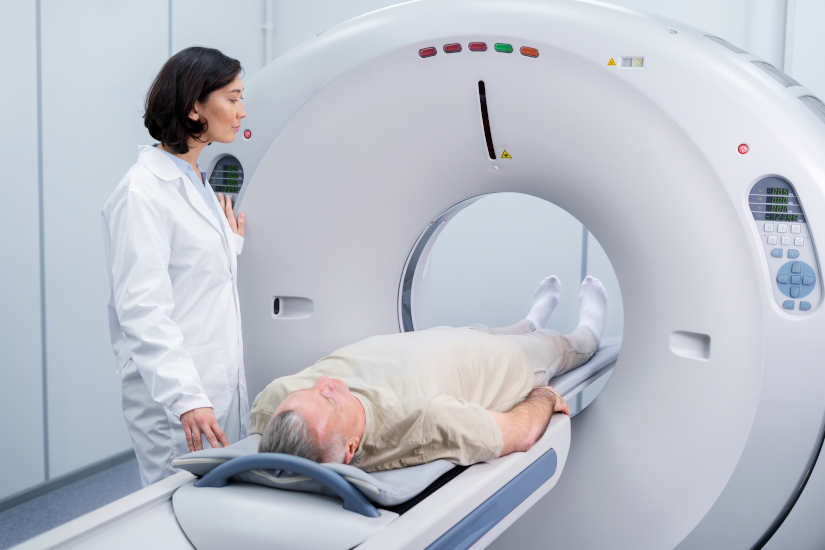
2. CT or computed tomography

It combines the X-ray technique together with the computed tomography or CAT scan to create cross-sectional images of the body. Subsequently, can be combined to generate a three dimensional image X-ray images. CT images are more detailed than those of a conventional X-ray and allow professionals to analyze the internal structures of the body from various angles.

Mammography

Breast radiography is used to detecting breast disorders, mainly breast cancer. Breast tissue is sensitive to radiation, so special mammography units are used to minimize radiation exposure. digital radiology equipment.

4. Fluoroscopy

X-rays and a fluorescent screen are used together. to obtain real-time images of the movement inside the body. It is also used to analyze diagnostic processes, such as following the path of a contrast agent.

One of the uses of fluoroscopy is to analyze heart movement and beats. For this, radiographic contrast agents are used to view the blood flow in the heart muscle, blood vessels and organs. This type of technique is also used to guiding an internally threaded catheter during cardiac angioplastya minimally invasive procedure to open clogged arteries that supply blood to the heart.

5. Therapeutic use of radiotherapy for the treatment of cancer.

Another use of X-rays is as therapeutic technique to destroy tumors and cancer cells. The dose of radiation used to treat cancer is higher than the radiation used in diagnostic tests. This type of therapeutic radiation can come from X-ray equipment or radioactive material. that is placed in the body or bloodstream.

Types of X-ray machines

What types of X-ray machines are available on the market? We can differentiate the following medical equipment using this technology:

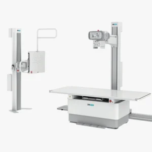
Conventional X-ray machines

They are the most basic equipment and are designed to obtain static images of the internal structures of the body. It is used for diagnose bone fractures, pulmonary evaluation by means of a chest X-ray and the identification of dental problems.

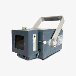
Portable X-ray machines

This type of X-ray machines are lightweight, compact and portableThe products can be easily transported. Used in emergencies and rural areasand to care for patients who cannot be transferred.

Digital X-ray machines

They are replacing film plates with digital detectors to develop a real-time diagnostics and the generated images have a high resolution and are of higher quality.

Fluoroscopy systems

These are specific devices that use X-ray technology for the following purposes observing dynamic processes in the body in real time. These machines are used for minimally invasive surgical procedures, gastrointestinal studies and orthopedic diagnostics.

Mammography machines

They are designed for perform breast tissue studies. They are essential for the screening for tumors, abnormalities and breast cancer. In this case, the X-ray emission is low energy in order to better analyze the soft tissues that make up the breasts.

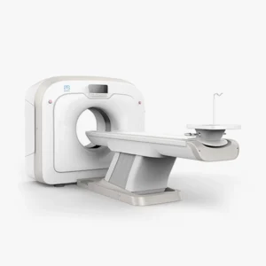
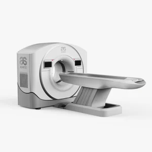
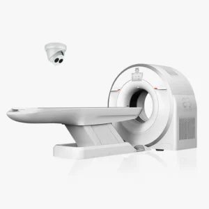
Computed tomography or CT equipment

These units are designed with a advanced system that uses X-rays to create detailed, three-dimensional images of the body. It is highly accurate and is used to evaluate internal injuries, tumors, as well as brain, thoracic, abdominal and extremity studies.

C-arc

These X-ray machines are equipped with a C-shaped arm que emits the X-rays from one end and captures digital images at the other end. C-arc is used for image-guided surgical procedures and in orthopedic and cardiovascular interventions. It offers a deeper analysis, since the area to be analyzed can be visualized from different angles.

Dental X-ray machines

This type of device is designed for capture images of the teeth and various maxillofacial structures. On the one hand, there are the intraoral equipment which capture images of the inside of the mouth and, on the other hand, there are the extraoral equipment which include panoramic systems that take full images of the jaw and mouth. They are mainly used for the diagnosis of caries, periodontal diseases and orthodontic planning.

X-ray machines for bone densitometry

X-rays are used to measuring bone mineral densityand, therefore used to diagnose osteoporosis and perform the follow-up of bone loss treatment.


Meet our 4D Medical equipment


Conclusion

In conclusion, X-rays are a very complete technique that has a large number of uses in the health field and, depending on each medical need, there is specific X-ray equipment to analyze, study and treat various diseases.

If you are interested in purchase an X-ray machine for your clinic, health center or hospital, at 4D Médica we are specialists in the sale of radiological medical equipment and we offer an optimal after-sales service. Ask us about our equipment without obligation.


Contact 4D Médica


Bibliography

National Institute of Biomedical Imaging and Bioengineering (n. d.). X-Ray. Retrieved from: https://www.nibib.nih.gov/espanol/temas-cientificos/rayos-x

Nuclear Forum (n. d.). History of the first radiography. Retrieved from: https://www.foronuclear.org/actualidad/a-fondo/historia-de-la-primera-radiografia/

My Diagnosis (n. d.). Radiography: Definition and uses. Retrieved from: https://midiagnostico.es/radiografia-definicion-y-usos/

MSD Manual (n. d.). Radiographs. At Common imaging tests. Retrieved from: https://www.msdmanuals.com/es/hogar/temas-especiales/pruebas-de-diagn%C3%B3stico-por-la-imagen-habituales/radiograf%C3%ADas

Mayo Clinic (n. d.). X-Ray: About this test. Retrieved from: https://www.mayoclinic.org/es/tests-procedures/x-ray/about/pac-20395303

Kiko Ramos

CEO of 4D Médica. Expert in marketing and distribution of medical equipment.

How has DANA affected clinics and health centers?

How has DANA affected clinics and health centers?

Thousands of businesses affected, more than 200 deaths and severe damage to many families, businesses and local institutions. The passage of the DANA has had an impact on a total of 75 municipalities in the Valencian Community, two in Castilla-La Mancha and one in Andalusia. Three weeks later, the economic cost is estimated at 1,789 million euros, between repairs and economic reactivation measures, in addition to a further 53 million euros in losses due to inactivity.

According to the data of the evaluation report carried out by the Valencia Chamber of Commerce, the total impact was 1,843 million euros.. But how has it affected the healthcare sector and medical centers?

Retail stores, the main affected by the DANA

The stores and small businesses have been the main affected after the floods. Approximately, a total of 5,228 companies have been directly hit by the DANA, and more than 3,500 businesses have serious damage. For this reason, the government has earmarked a set of measures to repair the damage in the affected premises. The cost of all structural damage will amount to €145 million, in addition to the €394 million earmarked for the cleaning and replacement of assets125 million for the construction of the new plant, such as machinery and furniture, and inventory replenishment.

The healthcare sector and the medical centers concerned

In the healthcare sector, there are many medical, veterinary and dental clinics; physiotherapy centers and health centers which have suffered numerous damages. Floods have affected a multitude of diagnostic imaging and laboratory equipment. and many of them could not be saved after the passage of the DANA.

4D Medica's services to help affected centers

In view of this situation, from 4D MedicaWe are doing our bit to help all the affected centers. Throughout the last few weeks, we have been carrying out different activities to technical assistance services free of charge. On the one hand, the removal of diagnostic equipment that have had serious mishaps and the reporting on the extent of the damages The companies can process the request for insurance indemnities and also the application for the different state aids.

Main solutions and measures for companies

The retail fabric has been the most affected, especially in the Valencian Community. An impact that could lead to the definitive closure of many businesses, as it is estimated that there is a potential risk that between 25% and 40% of commercial businesses will not reopen. For this reason, the government has promoted a series of economic reactivation measures to support businesses.

Industrial and service establishments have access to a subsidy of up to 7% of the value of the compensable damages36,896 euros. At the same time, it has been approved a 500 million to remove the accumulated sludge debris and repair water networks in the affected municipalities. On the other hand, the victims are exempted from paying the Real Estate Tax (IBI) and have a reduction in the Business Activity Tax (IAE) in 2024.

United to restore clinics and health centers to normalcy

There is still much to be done, but together, we will help restore health and well-being. Many volunteers have volunteered to clean up and bring supplies and materials to the hardest hit areas. At 4D Medica, we are supporting all the companies and centers that need it. The aim is to enable the affected businesses in the healthcare sector to resume their activities.

Together, we will get things back to normal. If your center is one of those affected, you can contact us by the following means:

We will be happy to help you!

Bibliography

El País. (2024, November 12). These are the aids approved for 14,373 million for those affected by the DANA. Retrieved from: https://elpais.com/economia/2024-11-12/estas-son-las-ayudas-aprobadas-por-14373-millones-para-los-afectados-por-la-dana.html

Levante-EMV (November 12, 2024). The DANA in Valencia: economic impact on trade and agriculture. Retrieved from: https://www.levante-emv.com/economia/2024/11/12/dana-valencia-impacto-economico-comercio-111620058.html

What is a CT scan and what is it used for?

What is a CT scan and what is it used for?

The computed tomographyalso known as computed axial tomography, also known as computed tomography, or TAChas become one of the most popular techniques of image diagnosis most commonly used. It is a procedure that uses special X-ray equipment and advanced computers to obtain three-dimensional images with different slices of the body.

Since its clinical introduction in 1971, it has undergone successive advances that have allowed its application in different fields of medicine. At present, computed tomography is used to diagnose disorders such as cancer, cardiovascular conditions, infectious processes, trauma and diseases of the locomotor apparatus. In the following article, we analyze how it works, what it is used for and the origin and evolution of this diagnostic test.

How does a CAT scan work?

In order to perform this diagnostic imaging, the following is used computed axial tomography system which incorporates a X-ray scanners generating three-dimensional images with different cuts of the interior of the organism.

These slices are called tomographic images and are used for the following purposes study various internal regions of the bodyThe CT scanner can be used to view everything from organs, bones and soft tissues to blood vessels. In contrast to radiography, which only provides a two-dimensional representation, the CT scan makes it possible to observe the three-dimensional images. This makes it possible to analyze tissues with greater detail and clarity. Another aspect to note is that the CT scanner utilizes a X-ray source and has a ionizing radiation higher than that of an X-ray.

During the procedure, the CT scanner rotates around the circular opening of a threaded structure called Gantry. The patient lies on a bed and is inserted inside the scanner so that the specialist can analyze the tissues. The X-ray detectors are located in front of the X-ray source and generate a series of images through different cuts. Subsequently, are transmitted to a computer where the interior of the organism can be visualized and analyzed.

CT contrast medium

As with X-rays, dense structures within the body, such as bones, are easy to image. However, soft tissues are more difficult to image. For this reason, contrast media have been developed that increase the visibility of tissues during X-ray or CT scans. Contain a set of substances that are safe for patients and allow the X-rays to be stopped, so that the organs will be seen in greater detail in the test.

For example, to examine the circulatory system, an iodine-based intravenous contrast medium is injected into the bloodstream to illuminate the blood vessels.

What is CT used for?

CT is used as a clinical diagnostic test, in follow-up studies to analyze the patient's health status, in radiotherapy treatment planning, and even for screening asymptomatic individuals with specific risk factors. A computed tomography scan creates detailed images of the bodywhich include the brain, thorax, spine and abdomen.. Specifically, we can highlight the following uses:

  • To help diagnose the presence of a cancer or tumor.. It is one of the most widely used techniques to screen for the presence of colorectal cancer and lung cancer.
  • Obtain information about the stage of a cancer.
  • Determine if a cancer reacts to treatment.
  • To detect the return or recurrence of a tumor.
  • Diagnose an infection.
  • Support technique to guide a biopsy procedure.
  • To guide some local treatmentssuch as cryotherapy, radiofrequency ablation and radioactive seed implantation.
  • Radiotherapy planning external beam or surgery.
  • Study the blood vessels.

When did computed tomography come into being?

Computed tomography was introduced in 1971 as an X-ray modality. which allowed axial images of the brain to be obtained, so it was a clinical method used specifically in the neuroradiology area. Its evolution has made CT a versatile imaging technique with which three-dimensional images of any anatomical area can be obtained. Currently, it is a diagnostic imaging equipment with a wide range of diagnostic capabilities. wide range of medical applications in oncology, vascular radiology, cardiology, traumatology or interventional radiology.

Evolution: From its beginnings to the present day

At 1971The following were developed first CT scanners for clinical use. During these early years, the EMI-scanner was used, with which brain data could be obtained and the calculation time per image was about 7 minutes in total. Soon after, scanners applicable to any part of the body were developed. At 1973In the early 1990s, the axial scannerswhose equipment had only one single row of X-ray detectors. Subsequently, it was when the helical or spiral scannerswhich incorporated multiple detector rowsits clinical use had a significant impact on the widely used and are the ones currently in use.

Current CT equipment: Main improvements and types

The evolution of the medical equipment has made it possible to obtain significant improvements. In today's systems, the image quality and offer both a better quality of life and a better spatial resolution as a low contrast resolution. In addition, nowadays, the following are also available CT scanners designed for specific clinical applications. Among them, we can highlight:

  • Specific CT equipment for radiotherapy treatment planning: These scanners offer a larger aperture diameter than usual, thus allowing a study with a wider field of view. Thus, the images generated have greater detail and clarity.
  • Hybrid equipment integrating CT scanners with other imaging techniquesHybrid solutions are now available. Among them, we can highlight the CT scanner incorporating a positron emission tomograph (PET) or a single photon emission tomograph (SPECT).
  • Specialized scanners for new indications in diagnostic imagingDual-source" CT scanners, which are equipped with two X-ray tubes, have been developed, as well as "volumetric" CT scanners, which incorporate up to 320 detector rows, making it possible to obtain complete data on the organs analyzed in a single use.

Meet our 4D Medical equipment

Main risks

CT scans can diagnose serious diseases and conditions such as cancer, hemorrhage or blood clots. An early diagnosis is essential in order to find a solution as soon as possible and save lives. However, it is true that it is a test that presents some risks that are important to discuss:

X-Ray

One of the main risks of CT is that it utilizes the X-rayswhich produce ionizing radiation. This type of radiation can have certain effects on the organism and it is a risk that increases with the number of exposures to which a person is subjected. However, the risk of developing cancer by the radiation emitted by the X-rays is generally low.

Use in pregnant women and children

In the case of pregnant women, there are no risks for the baby if the area of the body being imaged is not the abdomen or pelvis. But, medical professionals often perform tests that do not use radiation, such as the magnetic resonance imaging or ultrasound. As for the childrenare more sensitive to ionizing radiationas they have a longer life expectancy and the risk of developing cancer may be higher compared to adults.

Reactions to contrast medium

On the other hand, another aspect to be highlighted is that some patients may have allergic reactions to contrast medium and, in very specific cases, temporary renal insufficiency. In this situation, intravenous contrast media should not be administered to patients with abnormal renal function.


Conclusion

As we have been able to analyze, computed tomography or CT is very useful for detailed and precise analysis of certain internal tissues and organs. By means of X-rays, certain conditions or serious diseases can be studied, which is why it is essential for clinical diagnosis and its application in different fields of medicine.

Are you interested in a CAT scanner? Contact us and we will advise you without obligation so that you can choose the most suitable medical equipment for your clinic or hospital.

Contact 4D


Bibliography

International Atomic Energy Agency (n.d.). Computed tomography (CT). Retrieved from https://www.iaea.org/es/recursos/proteccion-radiologica-de-los-pacientes/informacion-para-los-pacientes-y-la-poblacion/tac

National Cancer Institute (n.d.). Computed Tomography (CT): Fact Sheet. Retrieved from https://www.cancer.gov/espanol/cancer/diagnostico-estadificacion/hoja-informativa-tomografia-computarizada

National Institute of Biomedical Imaging and Bioengineering (n.d.). Computed tomography (CT). Retrieved from https://www.nibib.nih.gov/espanol/temas-cientificos/tomograf%C3%ADa-computarizada-tc

MSD Manual (n.d.). Computed tomography (CT). Retrieved from https://www.msdmanuals.com/es/hogar/temas-especiales/pruebas-de-diagn%C3%B3stico-por-la-imagen-habituales/tomograf%C3%ADa-computarizada-tc?ruleredirectid=756#M%C3%A1s-informaci%C3%B3n_v21423499_es

Bernabéu, J. L., Bueno, E., & Figueroa, J. (2016). The use of computed tomography in medical physics. Journal of Medical Physics, 17(2), 125-133. Retrieved from https://revistadefisicamedica.es/index.php/rfm/article/view/115/115

MedlinePlus (n.d.). Computed tomography. U.S. National Library of Medicine. Retrieved from https://medlineplus.gov/spanish/ency/article/003330.htm

Kiko Ramos

CEO of 4D Médica. Expert in marketing and distribution of medical equipment.

Asturian Healthcare receives 54 state-of-the-art ultrasound scanners from DiagXimag

Asturian Healthcare receives 54 state-of-the-art ultrasound scanners from DiagXimag

The Principality of Asturias Health Service (Sespa) has received 54 state-of-the-art ultrasound scanners 1.7 million. Following the award in April 2024 of the public procurement contract for the DiagximagOn September 30, the delivery of the equipment was signed. The ultrasound scanners, which were delivered during the months of July and August, represent a very important step forward in the modernization and digitalization of the healthcare sector in the Asturian region. 

How are Diagximag's state-of-the-art ultrasound scanners different?

Diagximag, a subsidiary of Diagximag, an Asturian-based company 4D Medica and belonging to the group Substrate AIhas about state-of-the-art ultrasound scanners which offers a complete imaging solution. These Samsung-branded medical devices combine advanced technology and precision imaging of the different organs, tissues and internal structures of the body. It is an essential tool for diagnosing medical conditions, monitoring the health and development of the fetus during pregnancy, and for guiding certain medical procedures, such as biopsies and tissue extraction. Below, we analyze the main features that define Diagximag ultrasound scanners:

  • Equipment includes artificial intelligence and remote controlOne of the innovations of Diagximag ultrasound scanners is that they allow ultrasound scans to be performed using artificial intelligence. They differ in that they include the Sonosync function that allows radiologists to control the equipment completely remotely. In other words, from their own home, they can diagnose patients as if they were present at the medical center.
  • High image resolutionThey have a very good image resolution and incorporate Doppler technology, so that tissues and blood flow can be visualized with total clarity. This allows to visualize detailed images and perform a complete diagnosis of the area of the body to be analyzed.
  • Intuitive design for multiple uses in the clinical settingIn addition to their multiple functions, ultrasound scanners have an intuitive design that facilitates their use in different clinical environments. A fundamental aspect when it comes to increasing the efficiency of medical diagnostics.

What advantages and innovations do they offer in diagnostic imaging?

The ultrasounds are one of the most widely used medical techniques today, because it is a convenient, inexpensive, safe and non-invasive test. The medical devices used to perform this highly demanded test are ultrasound scanners. They have a rod-shaped toolcalled transducerwhich is responsible for detecting the waves produced inside the body. Through the use of a special gel which is applied to the skin of the area to be examined and the use of a computerThe images are displayed on the screen, which provide the information on fabrics.

The innovative technology based on the application of AI not only enhances the medical diagnostic experience, but also offers a major progress in telemedicine. In this way, the following can be realized rapid diagnostics no matter where the specialist is located. With this, it is possible to reach more regionsas there are many localities that do not have all medical services in health centers. The use of state-of-the-art ultrasound scanners means that more patients can receive a quick and accurate diagnosis, avoiding the need to travel to other regions that have more resources.

The use of these ultrasound scanners incorporating the latest technology provides an improvement in the health sector, so that now the Asturian region will be able to offer an effective and high quality diagnostic imaging, reducing efforts and limitations.

Latest entries

en_USEN
Resumen de privacidad

Esta web utiliza cookies para que podamos ofrecerte la mejor experiencia de usuario posible. La información de las cookies se almacena en tu navegador y realiza funciones tales como reconocerte cuando vuelves a nuestra web o ayudar a nuestro equipo a comprender qué secciones de la web encuentras más interesantes y útiles.