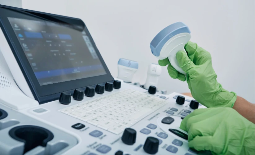
Ultrasound is a noninvasive medical technique that uses ultrasound to obtain real-time images of the inside of the body. The medical equipment used to perform an ultrasound scan is the ultrasound scannerwhich incorporates a device called a transducer. The ultrasound transducers are the main component of this medical equipment in the area of diagnostic imaging. They have the function of emitting high-frequency sound waves, which make it possible to observe the functioning and movements of the body's internal tissues and organs. Subsequently, they are responsible for generating the medical images that are displayed on the screen or monitor of the medical equipment, which are called sonograms.
The quality and usefulness of an ultrasound scan depend to a large extent on the transducer used. Therefore, in the following article, we discuss the operation of this device and provide a detailed guide to the different types of ultrasound transducers that exist. Do you want to know what their main advantages, functions and differences are? We will analyze them below!
Ultrasound transducers: Concept and operation
The transducer, also called ultrasound probeis the ultrasound component which converts electrical energy into sound waves, known as ultrasound. Its operation is based on the piezoelectric effect, a phenomenon in which certain crystals present in the transducer generate vibrations when receiving electric current, producing sound waves. In this way, the transducer or probe acts as a transmitter and receiver of
ultrasound.
When these waves penetrate the body and hit different structures and tissues, they return to the transducer in the form of echoes. Ultrasound scanners process this information and convert the captured ultrasounds into medical images that can be displayed on the equipment's screen. They are called sonograms and allow the following to be obtained visualize the functioning of the different tissues and organs in real time.
Use of transducers in ultrasound scanning
In the realization of a ultrasoundThe transducer plays a key role. The use of this device works as follows:
- Selection of the appropriate transducerThere are different types of transducers or ultrasound probes, so depending on the anatomical area to be evaluated, the physician or technician must select a specific transducer.
- Ultrasound gel applicationDuring an ultrasound scan, the transducer is coated with a conductive gel that slides over the patient's skin in the specific area to be analyzed. This gel eliminates the air between the skin and the transducer, which facilitates the transmission of the ultrasound waves and improves the quality of the images.
- Exploration of the area of interestThe transducer can be slid over the skin or inserted into a cavity in the case of transvaginal or transrectal ultrasound. While moving, the ultrasound scanner displays real-time images of the examined area on the screen.
- Parameter settingThe operator can modify certain parameters to improve image quality according to the depth and type of tissue to be analyzed. These include frequency, focus and gain.
- Image capture and interpretationSubsequently, the images generated are recorded for analysis and diagnosis, which creates an ultrasound scan that allows evaluation of the state of the organs and tissues.
Types of ultrasound transducers
Not all transducers perform the same function. Depending on the anatomical area to be analyzed, different resolutions and penetration depths are required. Therefore, a key aspect to increase diagnostic accuracy is to select the right transducers. transducers for ultrasound scanners adequate. To this end, it is important to to know the different options and models. Below, we provide a complete guide explaining the main types of transducers used in ultrasound along with their characteristics, advantages and clinical applications.
![]()
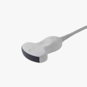
Linear transducers
Linear transducers are characterized by their rectangular shape and the emission of ultrasonic waves in parallel lines. They offer high resolution, but have lower penetration. They are mainly used for superficial studies in physiotherapy, podiatry and dermatology.
Advantages
- High image resolutionThis allows observation of fine anatomical details.
- Ideal for surface structuresThe frequency range is between 5 and 15 MHz.
- Excellent for vascular and musculoskeletal studies.
Clinical applications
- Vascular ultrasoundEvaluation of arteries and veins.
- Soft tissue ultrasoundThyroid, breast, muscle and joint examinations.
- Dermatological ultrasoundEvaluation of the skin and superficial structures.
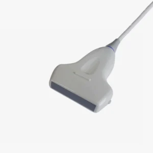
Convex or curvilinear transducers
These transducers have a curved shape that allows a larger field of view at intermediate and large depths. They generate sector or fan-shaped images. They have a greater penetration compared to the linear transducer. They are used for abdominal and gynecological studies.
Advantages
- Increased penetration than the linear transducer, includes frequencies between 2 and 6 MHz.
- Suitable for abdominal and pelvic studies.
- Has a wide image coverageIt is therefore very useful in large organ scans.
Clinical applications
- Abdominal ultrasoundEvaluation of the liver, kidneys, gallbladder and pancreas.
- Obstetric ultrasoundPregnancy monitoring and fetal assessment.
- Pelvic ultrasoundExploration and evaluation of the reproductive organs.
- Studies in pediatrics and general medicine.
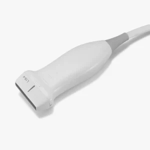
Sector or Phased Array Transducers
Sector transducers, also referred to as sector transducers, are phased arrayemit waves from a small spot. They emit waves in a narrow aperture scanning pattern and generate triangular or fan-shaped images. They have a high penetration, but have a lower resolution than linear transducers.
Advantages
- Allows scanning of deep structures without the need for extensive skin contact.
- Has a low frequency between 2 and 4 MHz, which guarantees excellent penetration.
- It is suitable for studies in confined spaces such as the thorax.
Clinical applications
- EchocardiographyEvaluation of the heart and large blood vessels.
- Pulmonary ultrasoundPulmonary parenchymal examination, diagnosis of thoracic pathologies and studies in intensive care.
- Emergency ultrasoundIt is used in FAST (Focused Assessment with Sonography for Trauma) studies in the area of trauma.
Endocavitary transducers (endovaginal and endorectal)
These transducers are designed to be inserted into body cavities and provide detailed, high-resolution images of internal organs at close range. This type of ultrasound probe is used in gynecology, obstetrics and urology specialties.
Advantages
- High image resolution due to its proximity to the organ to be examined.
- The frequency offered is intermediate-highThe resolution is between 5 and 9 MHz, thus offering a balance between resolution and penetration.
- Facilitates the detection of gynecological and prostate pathologies.
Clinical applications
- Transvaginal ultrasoundEvaluation of the uterus, ovaries and early pregnancy.
- Transrectal ultrasoundDiagnosis of prostate and rectal pathologies.
Microconvex transducers
This type of transducer is similar in design to convex transducers, but has a smaller surface area. Therefore, it is characterized by providing greater maneuverability in areas that are difficult to access. Among its different applications, microconvex transducers are used to perform examinations in pediatric patients, neonates and in the veterinary area.
Advantages
- Increased maneuverability in small anatomical areas.
- Intermediate frequency between 5 and 8 MHz, providing a balance between depth and resolution.
- It is the right choice for studies in patients difficult to explore with conventional transducers.
Clinical applications
- Pediatric and neonatal ultrasoundBrain and abdominal evaluation in neonates.
- Veterinary ultrasoundFor animal examinations.
- Studies in anesthesiology and intensive careIt is used as a guide for procedures such as catheter placement and punctures.
Volumetric transducers
These transducers generate three-dimensional images in real time using advanced technology with multiple piezoelectric crystals. They are used for 3D and 4D digital reconstruction to visualize anatomical volumes.
Advantages
- Detailed and volumetric images of anatomical structures.
- Allows evaluation of fetal morphology with greater precision.
- Enables navigation in advanced diagnostic studies.
Clinical applications
- Obstetric ultrasound in 3D and 4DDetailed evaluation of the fetus and detection of malformations and anomalies.
- Advanced gynecologic ultrasoundAccurate diagnosis of uterine and ovarian abnormalities.
- 4D EchocardiographyCardiac studies that allow the visualization of the heart in real time with high precision.
Special ultrasound transducers
In addition to conventional transducers, there are transducers designed for specific applications:
- Doppler transducersThey allow to evaluate blood flow in real time.
- Laparoscopic transducersMinimally invasive surgical procedures: They are used in minimally invasive surgical procedures.
- Array transducers or Matrix ArrayCapture multiple image planes simultaneously for more accurate reconstructions.
Meet our 4D Medical Ultrasound Transducers

Guide to choosing the right ultrasound transducer type
Selecting the right ultrasound transducer is essential to ensure high-quality images and accurate diagnoses. To do so, several aspects need to be considered:
Frequency
One of the key factors in the choice of transducer is the frequency, which is responsible for measuring the relationship between penetration depth and image resolution.. This is an essential aspect, as it determines its ability to penetrate the tissues and provide a clear image.
High frequency (greater than 7 MHz)
- Offers more detailed imagesbut with less penetration capacity.
- It is the ideal frequency for surface structures such as muscles, blood vessels and skin.
- Used in linear and endocavity transducers.
Low frequency (less than 5 MHz)
- Allows a increased penetration. However, its resolution is lower.
- It is used to evaluate deep organs such as the liver, kidneys and heart.
- It is located in convex and sector transducers.
If the objective is to study tissues close to the surface, as in a muscle ultrasound, a high-frequency transducer is recommended. On the other hand, to explore internal organs or structures located in deep areas, a low-frequency transducer should be chosen.
2. Specific clinical application
Before choosing a transducer, the following should be done take into account the medical specialty and the type of structures to be examined What types of transducers are recommended depending on the medical application?
Vascular and musculoskeletal ultrasound
It is recommended to use a linear transducerThe high-frequency imaging allows visualization of superficial structures such as arteries, veins, muscles and tendons in great detail.
Abdominal and obstetric examinations
Use a convex transducer to achieve greater penetration. It has a low frequency that allows deep penetration to evaluate organs such as the liver, kidneys and uterus.
Cardiac and pulmonary evaluation
Select a sector transducer (phased array). It can image the heart through confined spaces such as ribs and allows real-time dynamic studies to be developed.
Gynecology and urology
Choose a endocavitary transducer with high resolution. Its high frequency allows obtaining clear images of reproductive organs such as the uterus, ovaries and prostate.
Pediatrics and neonates
A microconvex transducer provides the best resolution to size ratio. Its smaller size facilitates scanning in infants and neonates.
Ultrasound in emergency and intensive care
You need a sector or microconvex transducer because of its portability and penetration capability for rapid imaging of critically ill patients.
Advanced 3D and 4D studies
It requires a volumetric transducer with three-dimensional reconstruction.
3. Necessary field of vision
The transducer design influences the coverage area of the ultrasound image. Depending on the size of the required field of view, the following options should be considered:
- For small and detailed structuresLinear or microconvex transducers are the best choice, as they provide high-resolution images in small areas such as blood vessels, muscles and joints.
- For studies of deep organs and large structuresIn this case, convex or sectorial transducers are recommended, since they allow visualization of large areas with good penetration. For this reason, they are the ones used in abdominal and cardiac studies.
4. Mobility and ease of use
In some clinical settings, portability and transducer size are other essential factors in obtaining a more efficient diagnosis.
- Studies in the operating room or emergency roomSectorial transducers are recommended, since their compact design and penetration capacity allow ultrasound scans to be performed in small spaces.
- General inquiriesConvex and linear transducers are the most commonly used due to their ease of use and versatility.
- Ultrasound-guided procedures (punctures, biopsies)Transducers with puncture guides are preferred to improve the accuracy of needle insertion.
| Transducer Type | Frequency (MHz) | Penetration Depth | Resolution | Main Applications |
|---|---|---|---|---|
| Linear | 5 – 15 | Download | High | Vascular, muscle, skin |
| Convex | 2 – 6 | Media | Media | Abdomen, obstetrics |
| Sectorial | 2 – 4 | High | Media | Cardiac, pulmonary |
| Endocavitary | 5 – 9 | Download | High | Gynecological, prostate |
| Microconvex | 5 – 8 | Media | Media | Pediatrics, anesthesia |
| 3D/4D | Variable | Variable | High | Obstetrics, cardiology |
Conclusion
The choice of transducer in ultrasound depends on the anatomical region to be evaluated and the level of detail required. From linear transducers for superficial structures to sectorial transducers for cardiac studies, each type of ultrasound probe has a specific function to optimize ultrasound diagnosis in various medical specialties.
Do you need more information? Contact us and the 4D Médica team will help you find the model that best suits the different needs of your clinic or medical center.
Bibliography
Borrego, R., & González Cortés, R. (2018).. Basic fundamentals of ultrasound. Spanish Society of Pediatric Intensive Care. Retrieved from https://secip.com/images/uploads/2018/09/1-FUNDAMENTOS-BASICOS-DE-ECOGRAF%C3%8DA.pdf
Pardell Peña, X. (2024). Ultrasonography and ultrasound. Authorea. Retrieved from https://www.authorea.com/doi/full/10.22541/au.172660489.98960333
DiagXimag(n.d.). Ultrasound and fluoroscopy specialists. Retrieved from https://diagximag.com/

Director of Diagximag. Distributor of medical imaging equipment and solutions.
![]()