The positron emission tomography (PET) scanning is the technique of diagnostic imaging more recent and modern. It is a nuclear medicine procedure that emerged in the 1970s in the United States and was introduced in Spain in 1995. To perform a positron emission tomography scan, a radioactive material, called a radiopharmaceutical, is administered intravenously and the diagnosis is then made using specific equipment: the PET scanner.
This medical device is equipped with a special camera that allows visualization of internal organs at the molecular and cellular leveloffering information on metabolic activity of the body's tissues. From the analysis of blood flow, oxygen consumption, glucose and protein metabolism, amino acid transport and cell division to the detection of biochemical changes.
In the PET technique, radioation is detected after administration of the radiopharmaceutical. To do this, you need a waiting time between 30 and 60 minutes for the substance to take effect and be distributed correctly throughout the patient's body. This diagnostic imaging test is used for to develop a metabolic study of the interior of the organismIt therefore provides a complement to the anatomical information offered by procedures such as computed tomography (CT) or magnetic resonance imaging (MRI).
One of the most recent advances in this area has been the development of hybrid equipment that combines two technologies in the same medical equipment. In 1998, the CT scanner began to be used in clinical practice. PET CTa device that incorporates the PET technique together with CT. A year earlier, in 1997, the hybrid PET MRI device was created by Mardsen and Cherry, which combines the anatomical images provided by MRI with the biochemical data from PET. However, it was not until 2009 that Phillips developed the first integrated system.
Currently, the use of positron emission tomography (PET) has made it possible to diagnose diseases in their earliest stages and, in turn, analyze the patient's response to specific treatments. Its ability to analyze functional changes before structural damage occurs in the body makes it key in the diagnosis and monitoring of multiple pathologies, especially in oncology, neurology and cardiology.
In the following article, we will analyze what this diagnostic technique consists of and the medical equipmentThe advantages and disadvantages, as well as their applications in clinical practice.
How does PET positron emission tomography work?
The positron emission tomography diagnosis consists of a process made up of different stages, which we analyze below:
Administration of the radiopharmaceutical
The first step in a PET study is the administration of a radioactive substancecalled radiopharmaceutical or radiotracer. This compound is generally introduced into the body intravenously, although in some cases it can be administered by inhalation or orally.
The most commonly used PET radiopharmaceutical is fluorodeoxyglucose (FDG). It consists of a glucose-like molecule that is labeled with fluorine-18, a radioactive isotope. The main reason for using FDG is that cells with high metabolic activity, such as cancer cells, consume more glucose than normal tissues. This allows the radiopharmaceutical to accumulate in areas of higher cellular metabolism, facilitating its detection.
2. Distribution and waiting
After administration of the radiopharmaceutical, the patient must remain at rest for 30 to 60 minutes for the substance to be adequately distributed throughout the body. During this time, it is recommended that the patient remain calm and avoid talking or moving excessively, since muscular activity could alter the uptake of the radiotracer and affect the quality of the images.
3. Patient positioning
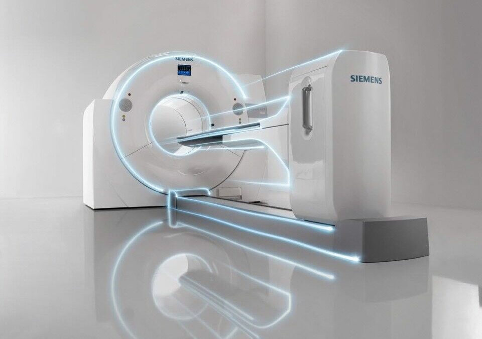
Once the radiopharmaceutical has been absorbed by the tissues, the patient is placed on a sliding stretcher which introduces it into the PET scanner.. This equipment consists of a ring of detectors that surrounds the patient and is capable of recording the radiation emitted by the radiopharmaceutical. The procedure has a duration between 15 and 45 minutesdepending on the type of study to be performed.
4. Diagnosis by PET scanner
The radiopharmaceutical injected into the patient emits positrons.which collide with the body's electrons, generating two gamma photons in opposite directions. The PET scanner detectors capture these gamma photons and record the exact location of each emission. Subsequently, the medical team is responsible for the reconstruction of a tomographic image detailed with the areas where the radiopharmaceutical has accumulated, reflecting the metabolic activity of tissues and organs.
5. Image processing and reconstruction
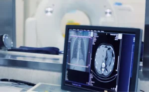
Once the data has been collected, specialized software processes the information and generates three-dimensional images of the distribution of the radiopharmaceutical in the patient's body. These images show the areas of increased metabolic activity (hyper uptake) in brighter colorswhile areas with lower metabolism appear in darker shades. This activity map allows physicians to accurately identify anomalies such as malignant tumors, neurodegenerative diseases or cardiac conditions.
6. Analysis and interpretation of results
Finally, specialists in radiology or nuclear medicine analyze the images. obtained to make a diagnosis. Depending on the case, the PET scan can be combined with other imaging techniquesas the computed tomography (CT) or the magnetic resonance imaging (MRI)as well as the use of hybrid equipment. This will provide a more complete view of the anatomy and function of the organs.
Source || Canva
Positron emission tomography advantages
Positron emission tomography (PET) is a highly advanced imaging technique with the following benefits:
Early detection of diseases
Allows you to identify metabolic abnormalities before visible structural changes occur in other imaging tests, which facilitates the early diagnosis of diseases. These include cancer, Alzheimer's disease and heart disease.
Real-time functional evaluation
In contrast to computed tomography (CT) or magnetic resonance imaging (MRI), which only analyze anatomy, PET provides information on how tissues and organs function at the cellular and molecular level.
Effective technique to detect cancer and metastases
PET is one of the most effective tools for the detection and localization of cancer and its metastasesThis allows us to know the extent of the disease and to plan the appropriate treatment.
Monitoring treatment response
This is a diagnostic technique used for evaluate how a patient is responding to chemotherapy, radiotherapy or immunotherapy treatments. In this way, it makes it possible to make adjustments to the therapeutic strategy in real time.
Combined technology for greater precision
The use of hybrid equipment allow both anatomical and functional information to be obtained at the same time. At present, PET-CT and PET-MRI equipment offer the most advanced benefits of using two techniques in a single study. Its use helps to improve diagnostic accuracy and radiation dose reduction received by the patient by up to 50 %.
Disadvantages of positron emission tomography
However, it also has a number of limitations that are important to analyze:
Exposure to ionizing radiation
The PET technique uses radioactive radiopharmaceuticals that expose the patient to ionizing radiation. Although its doses are low and safe, the amount of radiation increases significantly when using various diagnostic techniques.
High cost and limited availability
It is a expensive technique because of the need for specialized equipment and the use of radiopharmaceuticals. These substances require rapid distribution in order not to lose effectiveness. Therefore, one of their disadvantages is that they limit availability in certain hospitals and regions.
Waiting time and duration of the study
Before performing the PET scan, patient must wait 30 to 60 minutes after injection of the radiopharmaceutical. Thus, in comparison with other diagnostic techniques, waiting time increases the duration of the test.
Complex interpretation of images
Medical images obtained can be difficult to interpret.not all elevated glucose uptake indicates abnormalities. Therefore, alternative tests are required for a more accurate diagnosis.
Clinical uses and applications
Positron emission tomography is used in different medical specialties, specifically in oncology, neurology and cardiology. What are its main uses in clinical practice?
Oncology
- Early detection of malignant tumors.
- Identification of metastases and evaluation of cancer spread.
- Assessment of the response to treatment with chemotherapy or radiotherapy.
- Differentiation between benign and malignant tumors.
Neurology
- Early diagnosis of neurodegenerative diseases such as Alzheimer and Parkinson.
- Localization of epileptic foci in patients with treatment-resistant epilepsy.
- Evaluation of psychiatric illnesses and neurocognitive disorders.
Cardiology
- Determination of cardiac muscle viability in patients with myocardial infarction.
- Evaluation of blood flow and cardiac function in ischemic diseases.
Other medical applications
- Diagnosis of endocrine diseases, such as adrenal gland disorders.
- Detection of infections and chronic inflammatory diseases.
- Evaluation of gastrointestinal pathologies with metabolic involvement.
Conclusion
After analyzing the operation of positron emission tomography (PET), we can highlight that it is a fundamental tool in nuclear medicine to detect diseases in their early stages and evaluate the metabolic function of different organs and tissues.
Do you want more information about PET equipment? Contact us and we will offer you personalized advice to analyze the medical equipment you need in your clinic or hospital.
Bibliography
Acta Medica Group Angeles (2014). Use of Positron Emission Tomography (PET) in neurology. Retrieved from https://www.medigraphic.com/pdfs/actamedica/acm-2014/acm141i.pdf
Spanish Agency of Medicines and Health Products (AEMPS). (2007). PET Radiopharmaceuticals: Regulatory and Safety Considerations. Retrieved from https://www.aemps.gob.es/eu/publicaciones/articulo/docs/radiofarmacosPET_oct07.pdf
Spanish Society of Nuclear Medicine and Molecular Imaging (SEMNIM). (2019). Clinical applications of positron emission tomography. Retrieved from https://www.semnim.es/wp-content/uploads/2019/07/69.pdf
Author unknown (2018). Advances in the use of PET in oncology. Oncology (Spain), 27(8), 7-13. Retrieved from https://scielo.isciii.es/pdf/onco/v27n8/02.pdf
Carlos III University of Madrid (2002). Development and evaluation of radiopharmaceuticals in positron emission tomography. e-Archivo UC3M. Retrieved from https://e-archivo.uc3m.es/rest/api/core/bitstreams/e86f7bc6-a0e4-4b81-8886-7fbca138817b/content
Nuclear Safety Council (CSN). (n.d.). PET-RM: A revolutionary alliance. CSN. Retrieved from https://www.csn.es/-/pet-rm-una-revolucionaria-alianza#:~:text=El%20PET%2DRM%20naci%C3%B3%20en,desarroll%C3%B3%20el%20primer%20sistema%20integrado.

Director of Diagximag. Distributor of medical imaging equipment and solutions.
![]()