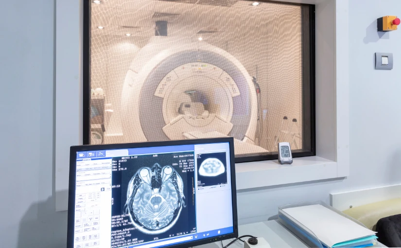
Customer service: 910 020 252 4dmedica@4dmedica.com
Classification of medical equipment according to risk

Classification of medical equipment according to risk

In the healthcare sector, patient safety comes first. For this reason, medical equipment must comply with certain regulations to ensure its reliability, efficiency and traceability. The classification of medical equipment regulatory requirements can be tailored according to the level of risk represented by each medical equipment for patients.

Depending on the type of riska series of controls and evaluations that must be carried out before they reach the market and can be marketed.. The higher the risk, the more rigorous and demanding the processes of clinical evaluation, quality control, technical documentation and post-marketing follow-up become. In the following article, we discuss how medical products are classified. in accordance with MDR (Medical Device Regulation).

Entry into force of the MDR standard for the classification of medical equipment

The MDR (Medical Device Regulation) regulation, officially known as the Regulation (EU) 2017/745 on medical devicesis the legal framework in force in the European Union for the regulation of medical devices. It is a European regulation that replaces the former Directive 93/42/EEC (MDD) and Directive 90/385/EEC on active implantable devices. In contrast to the directives, the MDR has a direct effect in all EU Member Stateswithout the need to adapt national laws.

It was approved on April 5, 2017 with the aim of reinforcing the safety, traceability and efficacy of these products on the European market, but its official entry into force did not arrive until May 25, 2017. As of May 26, 2021 and after a transition period of 4 years, its application became mandatory throughout the European Union.. For certain products certified under the old MDD standard, there is an extended transition period until 2027-2028.

Main changes in MDR regulations

The MDR replaced Directive 93/42/EEC (MDD). with a more precise and strict regulation. This regulation established new more detailed rules for classifying devices according to risk level. To this end, a number of specific criteria were applied, such as duration of use, invasiveness, area of the body affected and type of operation (active or passive). At the same time, it also added specific rules for medical software and the AI softwarewhich were not sufficiently covered before.

The main changes introduced were as follows:

  • More specific and strict classification rules, strengthening clinical and technical evaluation.
  • Increased control and new classification of implantable products and medical software
  • More rigorous assessment by notified bodies
  • Enhanced post-marketing surveillance requirements
  • Introduction of EUDAMED system for increased traceability and transparency
  • Direct and uniform application in all EU countries
  • Adapting the regulatory framework to new technologies, such as medical software and artificial intelligence applied to medicine

Classification of medical equipment according to MDR

The MDR regulation establishes a classification system in four different classes (I, IIa, IIb and III) according to the potential risk that the medical equipment represents for the user.

Class Risk Features Examples
Class I Under
  • Non-invasive
  • External or superficial use
  • No critical function
  • Bandages
  • Gloves
  • Simple thermometers
Class IIa Moderate
  • Short-term invasive
  • They may have software
  • Limited interaction with internal organs
  • Headphones
  • Short catheters
  • Basic medical software
Class IIb High
  • Invasive of medium/long duration
  • Act on vital functions
  • Prolonged use in internal organs
  • Respirators
  • Infusion pumps
  • Neonatal incubators
Class III Very high
  • Long-term implantable
  • Affect vital functions
  • Use in circulatory or nervous system
  • Pacemaker
  • Stents
  • Therapeutic AI software


Class I - Low risk

Class 1 medical devices are non-invasive equipment, for temporary or external usewhich do not interact directly with physiological functions critical body parts. Their design and use involves a minimal risk for the patient.

Main features

  • They do not require electricity or software to operate, they are "passive".
  • Used on the surface of the body or in a superficial manner
  • They may include variants such as:
    • Is (sterile)
    • Im (measuring function)
    • Go to (surgical reusables)

Examples

  • Gauze, bandages and sticks
  • Mercury-free thermometers
  • Non-sterile medical gloves
  • Manual wheelchairs

Type of evaluation

Generally, it is requires manufacturer's self-certificationexcept for the variants Is, Im and Ir, which require assessment by a notified body.

Class II - Moderate and high risk

Class 2 medical devices include two different modalities: Class IIa devices, which have a moderate risk, and Class IIb devices, which have a high risk.

Class IIa - Moderate risk

Includes short-term invasive medical deviceswhich are in use for less than 30 days, or active, which may have a moderate impact on health of the patient. This type of medical products can enter body cavities o use in non-critical diagnostic or therapeutic procedures.

Main features
  • Invasive through natural orifices or with limited medical intervention
  • Can be electrically powered or contain software
  • The risk is higher than in Class I, but still limited.
Examples
  • Hypodermic needles
  • Short-term catheters
  • Headphones
  • Non-critical monitoring software
Type of evaluation

Requires the participation of a notified body which evaluates technical documentation and clinical evidence, although it is less complex than in the higher classes.

Class IIb - High risk

It includes devices that can have a significant impact on vital physiological functions, which are long-term invasive or acting on critical internal organs. Also included is the software that directly influences relevant clinical decisions.

Main features
  • Invasives of medium or long duration
  • They act on the circulatory system or the central nervous system (if not for prolonged use)
  • Includes devices that deliver automatic treatments
Examples
  • Respirators
  • Neonatal incubators
  • Hemodialysis equipment
  • Diagnostic imaging software with AI
  • Programmable infusion pumps
Type of evaluation

Requires comprehensive clinical evaluation, technical review by a notified body and strict compliance with regulatory requirements.

Class III - Very high risk

Class 3 devices present the highest level of risk, since they may have a direct impact on vital functions or its use may involve critical intervention in the human body. Includes permanent implantable devices and stand-alone software for diagnosis or therapy.

Main features

  • Long-term or permanent implants
  • Long-term invasive devices in the central nervous system or circulatory system
  • Software with autonomous therapeutic functions

Examples

  • Pacemaker
  • Intracoronary stents
  • Cardiac valve prostheses
  • Brain implants
  • Artificial intelligence software provides oncology treatment solutions

Type of evaluation

Requires a mandatory full clinical evaluationincluding studies with patients. To this end, the notified body is involved at each stageThe following steps are required: development, manufacturing, documentation, post-sales surveillance. This type of medical equipment, being so high-risk, requires intensive post-marketing follow-up.

Factors that determine the classification of medical devices according to the MDR

The MDR regulation (Regulation (EU) 2017/745) establishes specific criteria for classifying medical devices according to their level of risk to the patient and the healthcare professional. What are the factors that determine the classification according to their risk?

Duration of use

This refers to the length of time the device remains in contact with the body. The longer the duration of contact, the greater the potential risk.

  • Temporary useLess than 60 minutes
  • Short-term useBetween 60 minutes and 30 days
  • Long-term use: More than 30 days

Degree of invasiveness

Evaluates whether and how the device penetrates the body. Implantable or surgical devices are rated higher.

  • Non-invasiveDoes not penetrate the body (e.g. bandages, external thermometers).
  • Invasive through natural orificesIt enters through the mouth, nose, ear, urethra, etc.
  • Surgical invasiveRequires medical intervention for insertion
  • ImplantableRemains inside the body for a prolonged period of time.

Affected body part

The MDR standard checks the site where the device acts in order to assess its risk. This risk increases when it affects a critical area of the human body.

  • Body surface or skin: lower risk
  • Internal organs or sterile cavitiesintermediate risk
  • Central nervous system or circulatory system: high risk

Active or passive use

Active devices can fail and their impact on the organism is greater, so they tend to be classified in higher classes.

  • Passive deviceOperates without energy source (e.g., syringes, dressings).
  • Active deviceRequires electrical or mechanical power to operate (e.g., respirators, infusion pumps).

Medical purpose

Another aspect that should be analyzed is the function performed by the device in the medical treatment or diagnosis. The higher the functional complexity and clinical relevance, the higher the risk in the classification. In this context, the following medical purposes can be differentiated:

  • Basic monitoring
  • Diagnose, treat or monitor medical conditions
  • Supports physiological functions
  • It is used for prevent diseases
  • Directly influences clinical decisions

Use of software

The MDR establishes clear rules for classifying medical software according to its use and clinical applications. The risk does not depend on the hardware, but on the purpose and clinical impact of the software.

  • Data management softwareClass I: Included in Class I
  • Software that aids in diagnostics or clinical decisionsClass IIa or IIb: Incorporated in Class IIa or IIb.
  • Autonomous software that makes therapeutic decisionsClass III: They are included in Class III because of increased risk.

Nature of the content covered

It is important to analyze whether the devices come into contact with the human body or alter the chemical composition of the organism. What options can we find depending on the nature of the content?

  • The device enters contact with blood, body fluids, or tissues
  • Modifies substances (chemically or thermally)
  • Administers medication or energy

The MDR regulation comprehensively analyzes how, where, how long and for what purpose a medical device is used. Each of these factors contributes to assigning it a risk class (I, IIa, IIb or III), which determines the legal and clinical requirements necessary to market it.

Importance of proper hazard classification of medical equipment

Classifying medical devices according to their risk is essential to ensure the safety of patients and users, and also to ensure that products comply with the appropriate regulatory requirements before they are marketed or used. What is the role of proper classification in the healthcare sector?

Protection of patients' health and lives

The classification makes it possible to identify the potential hazard level of a device. In this way, the necessary controls can be put in place to prevent failures that could cause harm to patients or healthcare professionals.

Determines the level of regulation and control

Higher-risk devices (Class III) require more rigorous clinical evaluations, testing, certification and post-market surveillance. In contrast, low-risk (Class I) devices follow simpler procedures, such as self-declaration of conformity by the manufacturer. This ensures that each device goes through a process commensurate with its level of risk.

Guidance to manufacturers and developers

Another of its functions is to assist manufacturers in understand key technical, clinical and documentary requirements that must be complied with according to the class of the device. Assessments and controls according to the risk of the medical equipment allow planning the process of development, validation, registration and market launch in an efficient and legally compliant manner..

Facilitates the work of health authorities

Regulatory authorities may prioritizing inspections and audits according to the risk associated with the product. This simplifies decision-making to authorize or restrict the use of certain devices.

Establishes trust in the marketplace and among users

Healthcare professionals and patients can be confident that they can rely on a product has been evaluated proportionally to the potential risks it represents. In this way, the promotes transparency, traceability and efficient management of incidents or product recalls.

It is a mandatory legal requirement

In most countries, classifying medical devices according to their risk is a legal requirement for approval and marketing (as in the European MDR Regulation, the FDA in the USA or the Chilean Institute of Public Health).

The classification of medical devices according to their risk is not only a regulatory procedure, but an essential tool to protect the health of patients and professionals, guarantee quality and make the entire healthcare system more efficient. If you work in the medical, technological or regulatory sector, knowing and applying this classification is the first step to ensure that your products reach the market in a safe, legal and responsible way.

Bibliography

Eurofins (n.?f.). What is a medical device/medical device? Eurofins Spain. Retrieved March 27, 2025, from https://www.eurofins.es/consumer-product-testing/industrias/productos-sanitarios/que-es-un-dispositivo-medico-producto-sanitario/

DQS Global (n.?f.). Understand the classification of medical devices according to the EU Medical Devices Regulation. Retrieved March 27, 2025, from https://www.dqsglobal.com/es-es/formacion/blog/comprender-la-clasificacion-de-los-productos-sanitarios-con-arreglo-al-reglamento-sobre-productos-sanitarios-de-la-ue

European Commission (2021). Medical Devices - Sector. European Commission - Public Health. Retrieved March 27, 2025, from https://health.ec.europa.eu/medical-devices-sector/overview_en

Public Health Institute of Chile (2019). Medical Device Hazard Classification Guidance. Retrieved March 27, 2025, from https://www.ispch.cl/sites/default/files/Guia_de_Clasificacion_de_Dispositivos_Medicos_Segun_riesgo_Formato_Institucional.pdf

Spanish Agency of Medicines and Health Products (s.?f.). Medical devices. Government of Spain - AEMPS. Retrieved March 27, 2025, from https://www.aemps.gob.es/productos-sanitarios/

European Union (2017). Regulation (EU) 2017/745 of the European Parliament and of the Council of 5 April 2017 on medical devices.. Official Journal of the European Union. Retrieved from https://eur-lex.europa.eu/legal-content/EN/TXT/?uri=CELEX%3A32017R0745

Luis Daniel Fernandez Perez

Director of Diagximag. Distributor of medical imaging equipment and solutions.

4D Médica provides medical equipment to the CRAS in Burgos and Valladolid

4D Médica provides medical equipment to the CRAS in Burgos and Valladolid

4D Médica has collaborated with the Burgos and Valladolid Wild Animal Recovery Centersproviding medical equipment in the area of image diagnosis in the field of veterinary radiology. At present, in Castilla y León, there are three Wild Animal Recovery Centers (CRAS) in the provinces of Valladolid, Burgos and Segovia and two Wild Animal Reception Centers (CRF) in the provinces of Zamora and Salamanca. All of them are part of the network of wildlife care centers of the Natural Heritage Foundation of Castilla y León.

The CRAS have the primary function is to care for and recover injured wildlife species. with the aim of rehabilitating them and reintroducing them into their natural habitat. According to the Regional Ministry of the Environment, the last year has closed with record figures in the CRAS. Throughout 2024, a total of 8,600 specimens have been cared for in the network, an increase of 22%.

Among the different CRAS of the Community, Burgos and Valladolid are the provinces with the highest percentages of specimens received. According to data from the Junta de Castilla y León, Burgos has a total of 1,402 animals attended and Valladolid has 1,376 animals, representing 28% and 21% of the entries respectively.

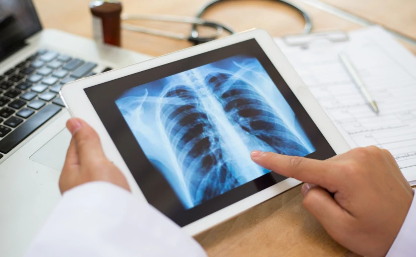
The collaboration between 4D Médica and the animal recovery centers in Burgos and Valladolid has arisen through a public bidding by the Junta de Castilla y León (Castilla y León Regional Government). In this project, we have supplied different X-ray equipmentas well as protective equipment for professionals in the veterinary field.

The Burgos and Valladolid Wild Animal Recovery Centers

The Wild Animal Recuperation Centers in Burgos and Valladolid are managed by the Junta de Castilla y León and play a fundamental role in the wildlife protection, conservation and recovery. They are responsible for the care and rehabilitation of injured, sick or vulnerable wildlife, to later reintroduce them into their natural habitat.

The CRAS de Burgos was inaugurated on March 25, 2015. and is located in the town of Albillos, Burgos. Its facilities have more than 16,300 m² within a 47,000 m² plot of land and allow it to take care of a total of 100 birds and 25 mammals simultaneously. It is the largest recovery center in the Community of Castilla y León, making it a reference point for the provinces of Burgos, Palencia and Soria. For its part, the Wild Animal Recovery Center of Valladolid has been working with wildlife since 1989..

What are the main functions of recovery centers?

The CRAS of Burgos and Valladolid play an essential role in the specialized care of wildlife. In turn, they collaborate with institutions and scientific research projects on environmental and biodiversity conservation in the region of Castilla y León. Specifically, they perform the following functions:

Reception, veterinary care and rehabilitation of wild animals

Both centers receive wild animals from citizens, environmental agents or law enforcement agencies, where they undergo a thorough examination of the animals. veterinary diagnosis to identify injuries, wounds or diseases. Once at the center, the animal recovery in order to rehabilitate them and reintroduce them into their natural habitat in the shortest possible time, whenever feasible.

Breeding in captivity of protected or endangered species

They also participate in captive species breeding programs to reinforce wild populations or endangered animal species.

Biodiversity conservation

Contribute to the maintenance and recovery of native species and disease control which can affect wildlife, livestock and the human population, known as zoonoses.

Scientific research and health surveillance

Another of its functions is to collect data on the animals treated and deceased. From the different causes of admission, injuries and pathologies to the study of the evolution of the disease. In the Valladolid and Burgos centers, the following are carried out anatomopathological analysis of deceased specimensThis allows the detection of diseases, intoxications and other crimes against wildlife. These analyses also contribute to genetic censuses of protected species such as the Iberian wolf and the brown bear. At the same time, these centers participate in wildlife health surveillance studies and programs.

Environmental education and awareness

Through outreach activities, school visits or awareness-raising campaignspromote respect, respect for the environmental education and wildlife protection. Among their tasks, they are in charge of informing about the most common causes of entry of wild animals, such as roadkill, electrocution, illegal traps, etc.

Custody of species that cannot be reintroduced into the wild

Some animals cannot return to the wild by the presence of irreversible lesions. Thus, CRAS maintain and care for these animals. in the center and study the evolution of each one of them. for educational and scientific purposes.

Coordination with other entities

Recovery centers collaborate with the Territorial Environmental Service, police forces, research centers and other regional or state administrations. to protect wildlife and rehabilitate animals in need of veterinary care.

Medical equipment supplied by 4D Médica

In collaboration with the CRAS of Burgos and Valladolid, the following have been provided medical devices:

Two Digiray FireCR Spark X-Ray image acquisition systems

The FireCR Spark is a digital radiographic reading system developed by Digiray. This equipment is designed to provide high quality medical images in an efficient and adaptable way to various clinical needs.

Main characteristics:

  • High image qualityThe FireCR Spark uses advanced signal collection technology that ensures clear and detailed images, facilitating accurate diagnoses.
  • Compact and versatile designThe small size and light weight of the device allows it to be placed on a table or on the wall, optimizing space in clinical environments.
  • Support for multiple cassette sizesThe system supports a variety of cassette sizes to suit different imaging needs.
  • Adaptive processing speedThe FireCR Spark line offers models with different imaging speeds to meet the specific requirements of veterinary medical diagnostics.
  • Advanced softwareQuantorMed+ Imaging software is included, providing an intuitive and easy-to-use interface. It offers fast and efficient operation and unlimited software updates, allowing the system to be kept up to date with the latest enhancements and functionalities.

Neovet X-Ray Equipment

Neovet equipment is a veterinary radiology system developed by the manufacturer Sedecal. It is specifically designed to meet the imaging needs of veterinary clinics and hospitals. This medical equipment combines advanced medical technology with functionalities adapted to the veterinary environment, facilitating high quality imaging for a variety of animal species.

Main characteristics:

  • Versatility in different imaging modalitiesThe system offers both analog and digital solutions, adapting to the preferences and requirements of each clinic. In addition, it offers a choice of fixed or variable focal length and the possibility of angular projections, which expands the diagnostic options available.
  • Exclusive STII Software (Only 3 Simple Touches)This intuitive software enables optimal digital imaging with as few as three interactions, streamlining the image acquisition process and improving efficiency in the clinical workflow.
  • Control console optionsThe system offers three control console configurations to suit different spaces and preferences:
    • Embedded tube support
    • Pedestal
    • Fixed to the wall
  • Analog to digital upgrade kitFor clinics that initially opt for a conventional system, Sedecal offers an upgrade kit that converts the Neovet equipment to a digital system, allowing an easy transition to digital radiology and ensuring a good investment for the future.

Biochemistry equipment

A biochemistry team also called a biochemistry analyzer, is an automated device that can be used to analyze measures concentrations of chemicals in biological samples. Mainly blood and urine. These analyses provide information on the functioning of various organs and systems of the animalssuch as the liver, kidneys, pancreas or general metabolism.

Veterinary equipment is adapted to work with species-specific reference values, which is key in animals with very different physiologies. From dogs, cats, and horses to cattle or poultry.

Main functions in veterinary medicine

  • Evaluate the liver function.
  • Analyze the kidney function.
  • Detect metabolic disorders.
  • Electrolyte profile studywhere sodium, potassium, calcium and chlorine values are measured.
  • Follow-up of treatments or surgeries
  • Preventive control and routine check-ups

Hematology team

A hematology team is an automated analyzer that studies the cellular composition of animal blood.. By means of this device, it is possible to perform a complete hemogramadapted to the hematological particularities of different animal species. These devices use technologies such as electrical impedance, flow cytometry or colorimetry to count, classify and measure the characteristics of red blood cells, white blood cells and platelets.

Main functions in veterinary medicine

  • Diagnosis of hematological diseases
  • Follow-up of chronic or infectious diseases
  • Pre-surgical evaluation
  • Geriatric check-ups and preventive check-ups
  • Rapid evaluations in field environments

Leaded X-Ray protection equipment for veterinarians

The X-ray shielding equipment with plumbed for veterinarians have the function of protect veterinary personnel and auxiliaries from exposure to ionizing radiation. during the performance of radiographic studies on animals. This protective equipment consists of a set of garments and elements made of lead-containing materials, which are designed for block or attenuate X-raysthus protecting the most sensitive organs of the human body.

Although the doses used in veterinary radiology are usually low and localized, cumulative exposure can represent a significant risk if appropriate radioprotection in the clinical environment.

With this medical equipment provided by 4D Médica in the veterinary area, the CRAS of Burgos and Valladolid can improve diagnostic accuracy in the treatment, rehabilitation and reintegration of wild animals treated at both centers.

Bibliography

Natural Heritage Foundation of Castilla y León. (n.d.). Burgos Wild Animal Recovery Center. Retrieved from https://patrimonionatural.org/casas-del-parque/centros-de-recuperacion-de-animales-silvestres/c-r-a-s-burgos

Natural Heritage Foundation of Castilla y León. (n.d.). Valladolid Wild Animal Recovery Center. Retrieved from https://patrimonionatural.org/casas-del-parque/centros-de-recuperacion-de-animales-silvestres/c-r-a-s-valladolid

El Español (August 13, 2024). Castilla y León earmarks millions of euros to improve the network of wildlife recovery centers. Retrieved from https://www.elespanol.com/castilla-y-leon/sociedad/medio-ambiente/20240813/castilla-leon-destina-millones-euros-mejorar-red-centros-recuperacion-fauna-silvestre/877912528_0.html

Junta de Castilla y León. (December 27, 2023). The network of Wild Animal Recovery Centers of Castilla y León has managed almost 7,000 cases during 2022.. Retrieved from https://comunicacion.jcyl.es/web/jcyl/Comunicacion/es/Plantilla100Detalle/1284721258244/NotaPrensa/1285274200230/Comunicacion

Cadena SER (January 19, 2025). Wild bird hospitals in record numbers. Retrieved from https://cadenaser.com/castillayleon/2025/01/19/los-hospitales-de-aves-silvestres-en-numeros-record-radio-valladolid/

DIGIRAY Corporation (n.d.). FireCR Spark. Retrieved from https://m.digiray.co.kr/page/page11

SEDECAL. (n.d.). NEOVET. Retrieved from https://www.sedecal.com/es/productos/neovet/

Kiko Ramos

CEO of 4D Médica. Expert in marketing and distribution of medical equipment.

Emotional ultrasound: Getting to know the baby before birth is possible

Emotional ultrasound: Getting to know the baby before birth is possible

Can you imagine being able to see the baby's gestures during pregnancy? From how it moves and yawns, the moments when it opens its eyes, when it changes position and even how it plays with the umbilical cord. Knowing the baby before it is born and see all their real-time movements is possible through emotional ultrasound.

This is a type of ultrasound scan that goes from the beyond medical diagnosisoffering a more human and closer experience in the area of image diagnosis. Not only does it provide medical information, but it also plays a key role in strengthening the emotional bond between the parents and the baby during pregnancy and gestation.

Emotional ultrasound is one of the most innovative techniques in the field of ultrasound and, by means of ultrasound, the baby can be visualized in detail. The emotional ultrasound equipment that are used combine 3D technologywhich offers three-dimensional images, and 4D and 5D technology, which incorporates the movement of the fetus in real time with high image sharpness and quality. Therefore, emotional ultrasound not only ensures that the baby can be observed with a high resolution, but also that its gestures and movements can be seen in the mother's uterus.

In the following article, we discuss its main characteristics and differences with follow-up medical ultrasound scans, as well as all the advantages it offers.

Main features and advantages of emotional ultrasound

Emotional ultrasound provides added value by offering a more accurate closer experience of parents and family members with the babyWhat are its main characteristics and differences with a traditional medical ultrasound?

High image quality

Emotional ultrasounds use advanced technology that allows visualization of the fetus with high detail, generating sharp, moving images in real time.

Emotional bonding with the baby

The main objective of the ultrasounds is to check the correct development of the baby. However, the emotional ultrasound is a non-invasive test that provides a experience that is closer, more human and real for parents and family members. It is performed in a relaxing environmentThe nursery is equipped with soft music and adapted lighting to create a cozy environment. In some cases, family members are allowed to be present to share the moment and to be able to see the baby's movements and gestures.

In turn, the professionals who perform the ultrasound scan employ a warmer and closer approachexplaining every detail with sensitivity and empathy.

Complete monitoring of the fetus during pregnancy

In addition to visualizing the development of the baby in real time, emotional ultrasound is also used to perform a complete monitoring of the fetus during gestation. It is essential to analyze its neurophysiological statusas well as to detect possible anomalies and malformations.

Does not replace diagnostic medical ultrasound scans

It is important to keep in mind that, during pregnancy, follow-up medical ultrasounds should be performed. The emotional ultrasound does not replace in any case the different ultrasounds that the mother should have to evaluate the correct development and growth of the baby in the different stages of gestation. What are the different follow-up medical ultrasounds and when are they performed? We can distinguish the following:

  • Pregnancy confirmation ultrasound (week 6 and 8)It is developed to verify pregnancy and check that the embryo is in the mother's uterus.
  • First semester ultrasound scans (week 11 and 14)It is used to measure the length of the fetus, estimate the due date and check whether it is a single or multiple pregnancy.
  • Morphological ultrasound (week 18 and 22)It allows to examine in detail the organs and structures of the fetus. It helps to detect congenital malformations, evaluate natal growth and determine the sex of the baby.
  • Third trimester ultrasound (week 28 and 32)It is used to evaluate the baby's growth.
  • Prepartum ultrasound (36th and 40th week)This is the last ultrasound and is essential to check the baby's position, examine the approximate weight of the baby and the condition of the mother's amniotic fluid and placenta.

Use of ultrasound scanners with advanced technology

To perform the emotional ultrasound, specific medical equipment is used that incorporates the latest technology to visualize the baby in detail, with high image quality, sharpness and in real time. For this purpose, the following are used ultrasound scanners with advanced 4D and 5D technology.

Creation of memories in digital format

Emotional ultrasound not only offers real-time visualization of the baby, but also provides parents and family members to have a memory of that beautiful moment. The clinics that perform this type of 5D ultrasound provide the experience together with the delivery of the different ultrasound images and videos in digital format. so that parents can preserve, share and remember this beautiful memory of pregnancy and gestation.

When is it recommended to perform an emotional ultrasound?

Emotional ultrasound can be performed at any time during pregnancy. However, the most indicated time is between 25 and 30 weeks.since the baby is more developed and the baby's movements can be better visualized in the maternal uterus. But, it is important to keep in mind several factors to improve the visibility of emotional ultrasound:

  • Use of appropriate ultrasound scanners
  • Experience with this ultrasound modality by medical professionals.
  • Fetal position
  • Amount of amniotic fluid

What are the differences between 3D, 4D and 5D ultrasound?

3D, 4D and 5D ultrasounds are advanced ultrasound technologies that allow you to see the baby in real time with high detail. Although they are often confused, there are a number of differences between them.

Technology 3D Ultrasound 4D Ultrasound 5D Ultrasound
Definition Static image in three dimensions with greater anatomical detail. Real-time moving images with volume. High resolution images with realistic light and shadow effects.
Visualization Provides a still image of the fetus or internal structures. Displays live movements, such as gestures or heartbeats. More sharpness, texture and realism in the baby's features.
Utility Detection of malformations and anatomical studies. Evaluation of fetal movements and expressions. Hyper-realistic images for better diagnosis and emotional experience.
Image quality Good resolution with volume. Lower resolution due to real-time capture. High definition with light effects for greater realism.
Experience for parents Allows you to see the baby's facial features in a still image. It makes it easy to observe live movements, smiles and yawns. Ultra-detailed display with an almost photographic appearance.


3D Ultrasound

3D ultrasound is an advanced ultrasound technique that provides three-dimensional images of the fetus in the uterus. Unlike traditional 2D ultrasound, which only shows black and white slices in real time, 3D ultrasound reconstructs the image in depth, providing a detailed view of the baby and its facial features.

Advantages of 3D ultrasound

  • The baby's facial features and anatomy can be observed more accurately.both hands and feet.
  • Detection of congenital anomalies. Allows for a more detailed evaluation of bone structure, cleft lip and limb defects.
  • Emotional experience for parents. It provides a more realistic image of the baby, strengthening the bond with the parents-to-be.
  • Better visualization of fetal development. The various organs and tissues can be analyzed more precisely.

4D Ultrasound

4D ultrasound is an evolution of 3D ultrasound that adds real-time motion. It is based on the continuous capture of 3D images to generate the video effect. The three-dimensional moving images produced make it possible to show the baby's facial expressions live. Thus, the baby can be visualized gesturing, smiling, yawning or moving hands and legs.

Advantages of 4D ultrasound

  1. Allows parents and family members to see the baby on the move and in real time.
  2. Help to detect possible facial or body anomalies.
  3. Increases the emotional connection between the parents and the baby.

5D Ultrasound

5D ultrasound is an enhancement of 4D ultrasound, which provides higher quality images. It incorporates greater definition and realism in the baby's skin, has better illumination and contrast that allow the baby's skin to appear more natural and also includes a sense of depth and volume more detailed.

Advantages of 5D ultrasound

  • Sharper and more natural images.
  • Increased accuracy in facial feature recognition.
  • latest technology that offers a more realistic experience. It provides a more direct and closer contact of the parents with the baby.

Why offer emotional ultrasound in your clinic?

Knowing the baby before it is born is a unique moment for parents and family members. This establishes a closer, emotional and realistic connection with the baby. Moreover, it is not only the visualization of the fetus in motion, but provides a complete experience.

By offering emotional ultrasound in a clinic, parents can acquire the memory of one of the most beautiful moments of pregnancy: seeing the baby's gestures and movements in real time. This service not only generates a competitive differentiationIt also contributes to improving patient experience and increasing business profitability.

It is important to note that emotional ultrasound does not replace diagnostic medical ultrasound.but complements. While medical ultrasounds are necessary to evaluate the baby's state of health, emotional ultrasound offers a more detailed and aesthetic view of the fetus, without medical purposes.

For this purpose, the following are used state-of-the-art ultrasound scanners incorporating advanced 4D and 5D technology that allow you to see the baby with great clarity and in real time. In addition, they also help to control and monitor the baby's development, identifying possible risks and anomalies during pregnancy.

If you want to more information about ultrasound scanners to include this emotional ultrasound service in your clinic, do not hesitate to contact us. At DiagXimagWe are experts in ultrasound scanners and medical equipment sales, and we help you to choose the most appropriate ultrasound scanners according to the needs of your center.

Contact DiagXimag


Bibliography

Haute Autorité de Santé (2012). Physicalographies in medical and non-medical view: definitions and compatibility. Retrieved from https://www.cfef.org/archives/bricabrac/echoHAS.pdf

Hospital Quirónsalud Toledo (2023, October 27). Emotional ultrasound: the most advanced technology for the study of the fetus that reinforces emotional bonding with the baby. Retrieved from https://www.quironsalud.com/es/comunicacion/actualidad/ecografia-emocional-tecnologia-avanzada-estudio-feto-refuer

Luis Daniel Fernandez Perez

Director of Diagximag. Distributor of medical imaging equipment and solutions.

Interventional Radiology: Types, Advantages and Disadvantages

Interventional Radiology: Types, Advantages and Disadvantages

The interventional radiology (IR) is a specialized branch of the radiology area that combines techniques of diagnostic imaging with minimally invasive therapeutic procedures to diagnose and treat different diseases. In contrast to traditional surgical procedures, which require large incisions and long recovery times, interventional radiology allows for treat diseases without the need for open surgery. In this way, it stands out as an innovative discipline that reduces risks, recovery time and postoperative complications.

In the last decades, interventional radiology has experienced a great growth with the development of new technological advances both in imaging techniques and in medical equipment for the diagnosis and treatment of patients. interventional radiology. In the following article, we analyze what it consists of, its different types, as well as its main advantages and disadvantages.

What is interventional radiology?

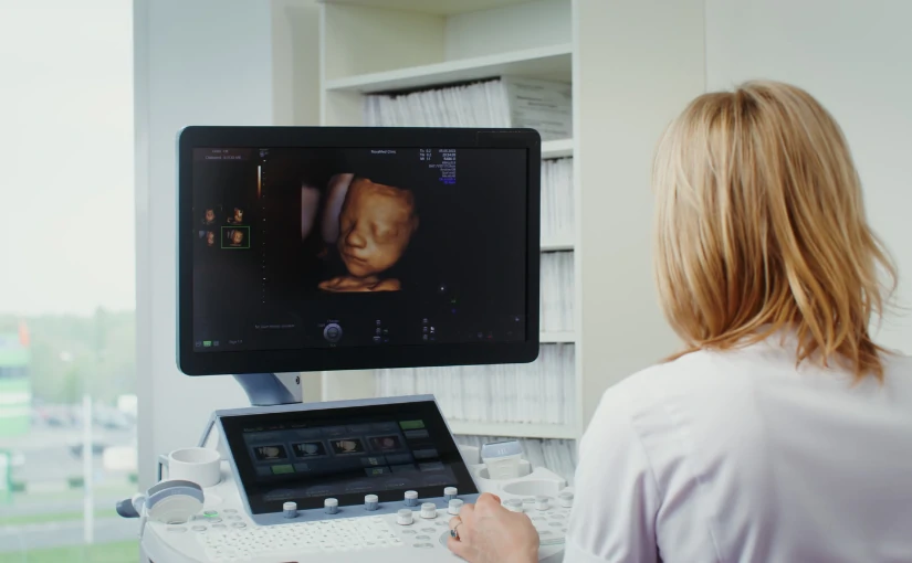
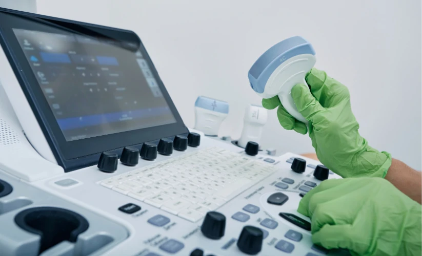
Interventional radiology uses a series of diagnostic imaging technologies to guide therapeutic procedures with a high precision. The main modalities used are the following X-raysthe ultrasoundsthe computed tomography (CT) scans and the magnetic resonance imaging (MRI).

These techniques offer detailed information on the patient's anatomy and physiology in real timeThis allows medical professionals to visualize specific areas of anatomical structures and access them by making small incisions. To do so, they use specialized instruments such as catheters and needles. The use of high quality images and live viewing capability during procedures not only facilitates device placement, but also plays a key role in minimizing the risks associated with the procedure and reducing damage to healthy tissues.

IR is a medical discipline that is used for the treatment of treatment of various medical specialtiesThe company's main areas of expertise include oncology, cardiology, neurology, vascular radiology and musculoskeletal medicine. In turn, it has the capacity to offer less invasive procedures for patients who have certain risks in conventional surgery, such as the elderly, as well as patients with advanced pathologies or with a high surgical risk.

Interventional radiology procedures are performed under the following conditions local anesthesia, patients are awake during the procedure. Therefore, the risks that may arise from the application of general anesthesia are reduced. Another aspect to highlight is that most procedures are performed on an outpatient basis. In this way, patients can return home on the same day of surgery, reducing hospital costs and increasing the efficiency of the healthcare system.

The multiple advances in technology offer a great projection in interventional radiology. The integration of artificial intelligence in medicine and robotics has a special relevance in this discipline, which will increase precision and efficiency in the treatment of many diseases.

Types of interventional radiology

Medical technology continues to advance and interventional radiology plays a fundamental role in modern medicine. It is now used in different medical specialties and covers a wide range of therapeutic procedures. The main types of interventional radiology include vascular, oncology, musculoskeletal, gastrointestinal, urological, thoracic and gynecological, which we discuss below:

Types of interventional radiology

Guided image diagnosis

One of the main functions of interventional radiology is the diagnosis of diseases through image-guided procedures. In many cases, it is necessary to take tissue samples or drain fluids accumulated in the body to obtain an accurate diagnosis. Through the use of imaging techniques, these procedures can be carried out with a millimetric precision and without the need for invasive surgery.

Main diagnostic procedures

  • Image-guided biopsiesBiopsies: Fine needles are used to remove tissue samples from organs such as the liver, lungs, thyroid or prostate. These biopsies make it possible to detect diseases such as cancer in its early stages.
  • Percutaneous drainageWhen there is fluid accumulation due to infection or inflammation, catheters are placed to remove it without the need for major surgery.
  • Puncture and aspiration of cysts or massesUsing a needle guided by ultrasound or tomography, physicians can remove cysts or reduce pressure in areas with fluid accumulation.

2. Vascular and endovascular treatment

The diseases of the circulatory systemsuch as arteriosclerosis, aneurysms and varicose veins can be effectively treated with interventional radiology techniques. In these cases, physicians use catheters and guidewires for accessing blood vessels and perform procedures that improve circulation or prevent serious complications. These treatments offer a less invasive alternative to conventional surgery, reducing hospitalization times and improving patients' quality of life.

Main treatments

  • Angioplasty and stentingIn patients with blocked arteries, a balloon is introduced through a catheter to widen the blood vessel. Subsequently, a stent, a small metal device that keeps the artery open and prevents future blockages, is placed.
  • Aneurysm embolizationWhen aneurysms, dangerous dilatations of the arteries, arise, micro-spirals or embolizing materials can be introduced to reduce the risk of rupture.
  • Treatment of varicose veins and vascular malformationsSclerotherapy techniques are used to close abnormal veins and improve circulation, eliminating aesthetic discomfort and associated circulatory problems.

Clinical applications

  • Peripheral arterial disease.
  • Cerebral and arterial aneurysms.
  • Stroke, cerebrovascular accident.
  • Varicose veins and venous malformations.

3. Interventional Oncology

In the field of oncology, interventional radiology has opened up new possibilities for the treatment of cancer. canceras it allows the localized destruction of tumorsThe impact on healthy tissues and its side effects are reduced. Therefore, interventional oncology represents an important effective and less aggressive alternative to surgery.

Procedures in interventional oncology

  • Percutaneous tumor ablationRadiofrequency, microwave or cryotherapy techniques are used to destroy tumors in the liver, kidney, lung and other organs without the need for open surgery.
  • Chemoembolization and radioembolizationChemotherapeutic drugs or radioactive particles are administered directly into the blood vessels that feed the tumor, reducing its size and preventing its growth.
  • Placement of catheters and central venous accesses.In patients requiring prolonged chemotherapy treatments, venous ports are inserted to administer the drugs more comfortably and safely.

Clinical applications

  • Liver, lung and kidney cancer.
  • Bone and soft tissue tumors.
  • Palliative treatment in oncology.

4. Traumatology and pain management

Interventional radiology procedures are also essential for the management of chronic pain and the treatment of musculoskeletal injuries. These procedures improve the quality of life for patients by reduce pain and restore joint function without the need to resort to open surgery.

Most common interventions

  • Joint infiltrations and nerve blocksAnesthetic and anti-inflammatory drugs are injected into joints such as the knee, hip or spine to relieve pain caused by arthritis or other conditions.
  • Cementoplasty (vertebroplasty and kyphoplasty)In this type of procedure, a bone cement is injected into fractured or osteoporosis-damaged vertebrae to reduce pain and improve spinal stability.
  • Aspiration of calcifications and drainage of articular cystsCalcium deposits in tendons or fluid accumulated in joints are eliminated, improving the patient's mobility and reducing pain.

Clinical applications:

  • Osteoporosis with vertebral fractures.
  • Herniated discs and chronic low back pain.
  • Rheumatoid arthritis and osteoarthritis.

5. Gastroenterology and urology

IR can be used to treat diseases of the digestive and urinary tract.

  • Placement of esophageal and biliary prosthesisStents are inserted in the esophagus, bile ducts or intestines to allow the passage of food or liquids in cases of obstructions caused by tumors.
  • Percutaneous nephrostomyDrainage: A drainage tube is inserted into the kidney to decompress urinary obstruction in patients with kidney stones or tumors.
  • Treatment of gastrointestinal bleedingEmbolization is used to stop bleeding from gastric ulcers or esophageal varices, avoiding emergency surgery.

Clinical applications in gastroenterology

  • Esophageal, liver and pancreatic cancer.
  • Hepatic cirrhosis with portal hypertension.
  • Biliary obstructions and intestinal strictures.

Clinical applications in urology

  • Urinary obstruction due to tumors or kidney stones.
  • Varicocele and fertility problems.
  • Benign prostatic hyperplasia.

6. Pulmonary and thoracic interventional radiology.

This specialty allows the diagnosis and treatment of thoracic diseases without the need for invasive surgical procedures.

Main procedures

  • CT-guided lung biopsyLung tissue sampling for cancer diagnosis.
  • Pleural drainage and pleurodesisElimination of fluid in the pleural space in cases of pleural effusion.
  • Embolization of pulmonary arteriovenous malformationsClosure of abnormal blood vessels in the lungs.

Clinical applications

  • Lung cancer and pleural diseases.
  • Recurrent pneumothorax.
  • Pulmonary vascular malformations.

7. Gynecology and obstetrics

In this medical specialty, the following treatments can be performed gynecological pathologies and pregnancy complications with image-guided procedures.

Main procedures

  • Uterine fibroid embolizationNon-surgical procedure that reduces the size of fibroids without removing the uterus.
  • Treatment of postpartum hemorrhageUterine arteries are occluded to stop severe bleeding after delivery.
  • Pelvic abscess drainageGynecological infections elimination with percutaneous catheters.

Clinical applications

  • Uterine fibroids and abnormal bleeding.
  • Severe postpartum hemorrhage.
  • Pelvic abscesses due to infections.

Advantages of interventional radiology

Interventional radiology offers many advantages and has transformed the treatment of many diseases, offering safer, less invasive procedures with shorter recovery times.

Minimally invasive procedures: Less risk and greater precision

One of the major advantages of interventional radiology is that it allows treatments to be performed without the need for open surgery. Instead of making large incisions, it uses small punctures in the skin through which catheters, microneedles and specialized devices are introduced.

This results in less damage to surrounding tissuesthere is a reduced risk of postoperative infections and you get a reduction of bleeding and scar formationimproving the patient's recovery.

Shorter hospital stay and faster recovery time

Interventional radiology procedures, being less aggressive to the body, allow the patient to recover in less time compared to conventional surgery. Many of the procedures are ambulatoryThe patient returns home after the procedure and is then hospital stay is reduced.

Another aspect to highlight is that the simpler and less invasive interventions. In this way, the decreases the consumption of painkillers since postoperative pain is less. At the same time, the patient can return to his or her daily activities and work in a shorter time, since the recovery times are shorter.

Less need for general anesthesia

Unlike traditional surgeries, which usually require general anesthesia, interventional radiology procedures are performed with local anesthesia and light sedation. This minimizes anesthetic risksespecially in patients with chronic diseases or advanced age. In addition to reducing the risk of complications, interventional radiology offers safer procedures for patients with cardiac or respiratory problems.

Highly accurate and efficient diagnosis and treatment

Interventional radiology uses real-time imaging to guide the placement of needles, catheters and other medical devices with extreme precision. The use of techniques such as fluoroscopy, ultrasound, computed tomography or magnetic resonance imaging offers different benefits:

  • Helps reduce the margin of error in complex procedures.
  • Increased success rate of oncology and vascular treatments.
  • Reduces collateral damage in adjacent structures.

Alternative treatment for patients who are not surgical candidates

For many patients with advanced disease or high surgical risks, interventional radiology is the only viable treatment option. It is a alternative for people suffering from advanced diseaseshave severe comorbidities or for those who reject invasive surgical procedures.

Covers multiple medical specialties

Interventional radiology is not limited to a single medical specialty, but is also encompasses several areas. Therefore, it provides a versatile treatment for treating diseases in different organs and systems, since it its approach is multidisciplinary. In addition, it is a discipline that is constantly evolving, thus allowing the application of new applications and technological improvements.

Lower cost compared to traditional surgeries

Although some interventional radiology procedures may involve more expensive medical equipment, their total cost is lower than that of conventional surgery. Among the main factors that reduce its costs, we can highlight the following aspects:

  • Reduced consumption of medical resources and hospitalization time.
  • Medication is reduced administered to patients.
  • Faster recovery.

Disadvantages of interventional radiology

Despite its many benefits, interventional radiology is not without its limitations and challenges. Although it represents a less invasive alternative to traditional surgery, there are factors that may limit its application or affect patient safety.

Limited availability and restricted access

One of the main challenges in interventional radiology is that not all hospitals and clinics have the necessary technology and trained specialists. to perform these procedures.

In rural areas or countries with fewer resources, patients may not have access to advanced imaging equipment or interventional radiologists, limiting the possibility of receiving these treatments. In these cases, patients will have to travel long distances to receive care, so many of them will have to opt for more invasive surgeries due to the unavailability of interventional radiology.

Another disadvantage is that not all health systems finance these procedures. and this may generate economic barriers that hinder access to this medical discipline.

Not all procedures are equally effective in the treatment of the disease.

Although interventional radiology offers effective solutions for many diseases, some procedures only control symptoms or slow disease progression, but do not completely eliminate the disease. Therefore, IR emerges as a temporary solution until the patient is able to undergo definitive treatment. On other occasions, some treatments must be repeated several times to increase its effectiveness.

Use of ionizing radiation in some procedures

Many interventional radiology procedures, especially those that utilize X-ray machines and fluoroscopy, expose the patient to ionizing radiation. Although the doses are usually low, repeated exposure may increase the risk to the patient.

What impact can it have on the patient? On the one hand, cumulative exposure over the years. could increase the risk of adverse effectsespecially in repeated procedures. In turn, in young patients or pregnant women, the risk-benefit ratio should be evaluated with caution.

Possible adverse effects and complications

Although interventional radiology is generally safer than surgery, it is not without risks and complications. As these are minimally invasive procedures, there is a possibility of adverse effects in certain patients:

  • Hemorrhage at the puncture siteMay occur in procedures that require the insertion of catheters in arteries or veins.
  • Allergic reactions to contrast mediumIn studies such as angiography and cholangiography, some patients may present severe allergic reactions to iodinated contrast material.
  • Infection at the puncture siteAlthough less frequent than in conventional surgeries, there is still a risk of infection.
  • Device migrationIn rare cases, a stent or embolization coil may dislodge and cause unwanted obstructions.

Recent discipline and limited availability of professionals

The success of interventional radiology depends largely on the skill and experience of the interventional radiologist. In contrast to traditional surgery, where surgeons have extensive experience, IR is a relatively new specialtyand, therefore, the availability of highly trained professionals is still limited.

Interventional radiology is a recent medical discipline that offers high precision, reducing the application of invasive treatments and open surgery. In recent years, it has had a great impact on modern medicine, improving the quality of life of patients and reducing postoperative complications, hospitalization times and health care costs.

Bibliography

Dalda Navarro, J. Á., Navarro Martín, M. T., Negre Ferrer, E., Negre Ferrer, C., Navarro Martín, A. B., & Dalda Navarro, V. (2024). Interventional radiology: minimally invasive treatments under image guidance. Health Research Journal, 5(6). Retrieved from https://dialnet.unirioja.es/servlet/articulo?codigo=9693488?

Lonjedo, E. (2019). Interventional radiology: as far as the image takes you. Annals (Reial Acadèmia de Medicina de la Comunitat Valenciana), (20). Retrieved from https://dialnet.unirioja.es/servlet/articulo?codigo=7710219

Kiko Ramos

CEO of 4D Médica. Expert in marketing and distribution of medical equipment.

Types of ultrasound transducers: Guide to choosing the right one

Types of ultrasound transducers: Guide to choosing the right one

Ultrasound is a noninvasive medical technique that uses ultrasound to obtain real-time images of the inside of the body. The medical equipment used to perform an ultrasound scan is the ultrasound scannerwhich incorporates a device called a transducer. The ultrasound transducers are the main component of this medical equipment in the area of diagnostic imaging. They have the function of emitting high-frequency sound waves, which make it possible to observe the functioning and movements of the body's internal tissues and organs. Subsequently, they are responsible for generating the medical images that are displayed on the screen or monitor of the medical equipment, which are called sonograms.

The quality and usefulness of an ultrasound scan depend to a large extent on the transducer used. Therefore, in the following article, we discuss the operation of this device and provide a detailed guide to the different types of ultrasound transducers that exist. Do you want to know what their main advantages, functions and differences are? We will analyze them below!

Ultrasound transducers: Concept and operation

Transducer, also called ultrasound probeis the ultrasound component which converts electrical energy into sound waves, known as ultrasound. Its operation is based on the piezoelectric effect, a phenomenon in which certain crystals present in the transducer generate vibrations when receiving electric current, producing sound waves. In this way, the transducer or probe acts as a transmitter and receiver of
ultrasound.

When these waves penetrate the body and hit different structures and tissues, they return to the transducer in the form of echoes. Ultrasound scanners process this information and convert the captured ultrasounds into medical images that can be displayed on the equipment's screen. They are called sonograms and allow the following to be obtained visualize the functioning of the different tissues and organs in real time.

Use of transducers in ultrasound scanning

In the realization of a ultrasoundThe transducer plays a key role. The use of this device works as follows:

  1. Selection of the appropriate transducerThere are different types of transducers or ultrasound probes, so depending on the anatomical area to be evaluated, the physician or technician must select a specific transducer.
  2. Ultrasound gel applicationDuring an ultrasound scan, the transducer is coated with a conductive gel that slides over the patient's skin in the specific area to be analyzed. This gel eliminates the air between the skin and the transducer, which facilitates the transmission of the ultrasound waves and improves the quality of the images.
  3. Exploration of the area of interestThe transducer can be slid over the skin or inserted into a cavity in the case of transvaginal or transrectal ultrasound. While moving, the ultrasound scanner displays real-time images of the examined area on the screen.
  4. Parameter settingThe operator can modify certain parameters to improve image quality according to the depth and type of tissue to be analyzed. These include frequency, focus and gain.
  5. Image capture and interpretationSubsequently, the images generated are recorded for analysis and diagnosis, which creates an ultrasound scan that allows evaluation of the state of the organs and tissues.

Types of ultrasound transducers

Not all transducers perform the same function. Depending on the anatomical area to be analyzed, different resolutions and penetration depths are required. Therefore, a key aspect to increase diagnostic accuracy is to select the right transducers. transducers for ultrasound scanners adequate. To this end, it is important to to know the different options and models. Below, we provide a complete guide explaining the main types of transducers used in ultrasound along with their characteristics, advantages and clinical applications.

Linear transducers

Linear transducers are characterized by their rectangular shape and the emission of ultrasonic waves in parallel lines. They offer high resolution, but have lower penetration. They are mainly used for superficial studies in physiotherapy, podiatry and dermatology.

Advantages

  • High image resolutionThis allows observation of fine anatomical details.
  • Ideal for surface structuresThe frequency range is between 5 and 15 MHz.
  • Excellent for vascular and musculoskeletal studies.

Clinical applications

  • Vascular ultrasoundEvaluation of arteries and veins.
  • Soft tissue ultrasoundThyroid, breast, muscle and joint examinations.
  • Dermatological ultrasoundEvaluation of the skin and superficial structures.

Convex or curvilinear transducers

These transducers have a curved shape that allows a larger field of view at intermediate and large depths. They generate sector or fan-shaped images. They have a greater penetration compared to the linear transducer. They are used for abdominal and gynecological studies.

Advantages

  • Increased penetration than the linear transducer, includes frequencies between 2 and 6 MHz.
  • Suitable for abdominal and pelvic studies.
  • Has a wide image coverageIt is therefore very useful in large organ scans.

Clinical applications

  • Abdominal ultrasoundEvaluation of the liver, kidneys, gallbladder and pancreas.
  • Obstetric ultrasoundPregnancy monitoring and fetal assessment.
  • Pelvic ultrasoundExploration and evaluation of the reproductive organs.
  • Studies in pediatrics and general medicine.

Sector or Phased Array Transducers

Sector transducers, also referred to as sector transducers, are phased arrayemit waves from a small spot. They emit waves in a narrow aperture scanning pattern and generate triangular or fan-shaped images. They have a high penetration, but have a lower resolution than linear transducers.

Advantages

  • Allows scanning of deep structures without the need for extensive skin contact.
  • Has a low frequency between 2 and 4 MHz, which guarantees excellent penetration.
  • It is suitable for studies in confined spaces such as the thorax.

Clinical applications

  • EchocardiographyEvaluation of the heart and large blood vessels.
  • Pulmonary ultrasoundPulmonary parenchymal examination, diagnosis of thoracic pathologies and studies in intensive care.
  • Emergency ultrasoundIt is used in FAST (Focused Assessment with Sonography for Trauma) studies in the area of trauma.

Endocavitary transducers (endovaginal and endorectal)

These transducers are designed to be inserted into body cavities and provide detailed, high-resolution images of internal organs at close range. This type of ultrasound probe is used in gynecology, obstetrics and urology specialties.

Advantages

  • High image resolution due to its proximity to the organ to be examined.
  • The frequency offered is intermediate-highThe resolution is between 5 and 9 MHz, thus offering a balance between resolution and penetration.
  • Facilitates the detection of gynecological and prostate pathologies.

Clinical applications

  • Transvaginal ultrasoundEvaluation of the uterus, ovaries and early pregnancy.
  • Transrectal ultrasoundDiagnosis of prostate and rectal pathologies.

Microconvex transducers

This type of transducer is similar in design to convex transducers, but has a smaller surface area. Therefore, it is characterized by providing greater maneuverability in areas that are difficult to access. Among its different applications, microconvex transducers are used to perform examinations in pediatric patients, neonates and in the veterinary area.

Advantages

  • Increased maneuverability in small anatomical areas.
  • Intermediate frequency between 5 and 8 MHz, providing a balance between depth and resolution.
  • It is the right choice for studies in patients difficult to explore with conventional transducers.

Clinical applications

  • Pediatric and neonatal ultrasoundBrain and abdominal evaluation in neonates.
  • Veterinary ultrasoundFor animal examinations.
  • Studies in anesthesiology and intensive careIt is used as a guide for procedures such as catheter placement and punctures.

Volumetric transducers

These transducers generate three-dimensional images in real time using advanced technology with multiple piezoelectric crystals. They are used for 3D and 4D digital reconstruction to visualize anatomical volumes.

Advantages

  • Detailed and volumetric images of anatomical structures.
  • Allows evaluation of fetal morphology with greater precision.
  • Enables navigation in advanced diagnostic studies.

Clinical applications

  • Obstetric ultrasound in 3D and 4DDetailed evaluation of the fetus and detection of malformations and anomalies.
  • Advanced gynecologic ultrasoundAccurate diagnosis of uterine and ovarian abnormalities.
  • 4D EchocardiographyCardiac studies that allow the visualization of the heart in real time with high precision.

Special ultrasound transducers

In addition to conventional transducers, there are transducers designed for specific applications:

  • Doppler transducersThey allow to evaluate blood flow in real time.
  • Laparoscopic transducersMinimally invasive surgical procedures: They are used in minimally invasive surgical procedures.
  • Array transducers or Matrix ArrayCapture multiple image planes simultaneously for more accurate reconstructions.

Guide to choosing the right ultrasound transducer type

Selecting the right ultrasound transducer is essential to ensure high-quality images and accurate diagnoses. To do so, several aspects need to be considered:

Frequency

One of the key factors in the choice of transducer is the frequency, which is responsible for measuring the relationship between penetration depth and image resolution.. This is an essential aspect, as it determines its ability to penetrate the tissues and provide a clear image.

High frequency (greater than 7 MHz)

  • Offers more detailed imagesbut with less penetration capacity.
  • It is the ideal frequency for surface structures such as muscles, blood vessels and skin.
  • Used in linear and endocavity transducers.

Low frequency (less than 5 MHz)

  • Allows a increased penetration. However, its resolution is lower.
  • It is used to evaluate deep organs such as the liver, kidneys and heart.
  • It is located in convex and sector transducers.

If the objective is to study tissues close to the surface, as in a muscle ultrasound, a high-frequency transducer is recommended. On the other hand, to explore internal organs or structures located in deep areas, a low-frequency transducer should be chosen.

2. Specific clinical application

Before choosing a transducer, the following should be done take into account the medical specialty and the type of structures to be examined What types of transducers are recommended depending on the medical application?

Vascular and musculoskeletal ultrasound

It is recommended to use a linear transducerThe high-frequency imaging allows visualization of superficial structures such as arteries, veins, muscles and tendons in great detail.

Abdominal and obstetric examinations

Use a convex transducer to achieve greater penetration. It has a low frequency that allows deep penetration to evaluate organs such as the liver, kidneys and uterus.

Cardiac and pulmonary evaluation

Select a sector transducer (phased array). It can image the heart through confined spaces such as ribs and allows real-time dynamic studies to be developed.

Gynecology and urology

Choose a endocavitary transducer with high resolution. Its high frequency allows obtaining clear images of reproductive organs such as the uterus, ovaries and prostate.

Pediatrics and neonates

A microconvex transducer provides the best resolution to size ratio. Its smaller size facilitates scanning in infants and neonates.

Ultrasound in emergency and intensive care

You need a sector or microconvex transducer because of its portability and penetration capability for rapid imaging of critically ill patients.

Advanced 3D and 4D studies

It requires a volumetric transducer with three-dimensional reconstruction.

3. Necessary field of vision

The transducer design influences the coverage area of the ultrasound image. Depending on the size of the required field of view, the following options should be considered:

  • For small and detailed structuresLinear or microconvex transducers are the best choice, as they provide high-resolution images in small areas such as blood vessels, muscles and joints.
  • For studies of deep organs and large structuresIn this case, convex or sectorial transducers are recommended, since they allow visualization of large areas with good penetration. For this reason, they are the ones used in abdominal and cardiac studies.

4. Mobility and ease of use

In some clinical settings, portability and transducer size are other essential factors in obtaining a more efficient diagnosis.

  • Studies in the operating room or emergency roomSectorial transducers are recommended, since their compact design and penetration capacity allow ultrasound scans to be performed in small spaces.
  • General inquiriesConvex and linear transducers are the most commonly used due to their ease of use and versatility.
  • Ultrasound-guided procedures (punctures, biopsies)Transducers with puncture guides are preferred to improve the accuracy of needle insertion.


Transducer Type Frequency (MHz) Penetration Depth Resolution Main Applications
Linear 5 – 15 Download High Vascular, muscle, skin
Convex 2 – 6 Media Media Abdomen, obstetrics
Sectorial 2 – 4 High Media Cardiac, pulmonary
Endocavitary 5 – 9 Download High Gynecological, prostate
Microconvex 5 – 8 Media Media Pediatrics, anesthesia
3D/4D Variable Variable High Obstetrics, cardiology


At DiagXimagAs experts in the sale of ultrasound scanners and medical equipment, you can find a wide range of ultrasound scanners and transducers to suit the different needs of your clinic or medical center.

The choice of transducer in ultrasound depends on the anatomical region to be evaluated and the level of detail required. From linear transducers for superficial structures to sectorial transducers for cardiac studies, each type of ultrasound probe has a specific function to optimize ultrasound diagnosis in various medical specialties.

Contact DiagXimag

Bibliography

Díaz-Rodríguez, N., Garrido-Chamorro, R. P., & Castellano-Alarcón, J. (2007).. Methodology and techniques. Ultrasound: physical principles, ultrasound scanners and ultrasound language. Family Medicine. SEMERGEN, 33(7), 362-369. Retrieved from https://www.elsevier.es/es-revista-medicina-familia-semergen-40-articulo-metodologia-tecnicas-ecografia-principios-fisicos-13109445

Borrego, R., & González Cortés, R. (2018).. Basic fundamentals of ultrasound. Spanish Society of Pediatric Intensive Care. Retrieved from https://secip.com/images/uploads/2018/09/1-FUNDAMENTOS-BASICOS-DE-ECOGRAF%C3%8DA.pdf

Pardell Peña, X. (2024). Ultrasonography and ultrasound. Authorea. Retrieved from https://www.authorea.com/doi/full/10.22541/au.172660489.98960333

DiagXimag(n.d.). Ultrasound and fluoroscopy specialists. Retrieved from https://diagximag.com/

Luis Daniel Fernandez Perez

Director of Diagximag. Distributor of medical imaging equipment and solutions.

en_USEN
Resumen de privacidad

Esta web utiliza cookies para que podamos ofrecerte la mejor experiencia de usuario posible. La información de las cookies se almacena en tu navegador y realiza funciones tales como reconocerte cuando vuelves a nuestra web o ayudar a nuestro equipo a comprender qué secciones de la web encuentras más interesantes y útiles.